info@gucluhanguclu.com
Beşyol, Florya, Akasya St. No:4 Apt:1, 34295 Küçükçekmece/Istanbul
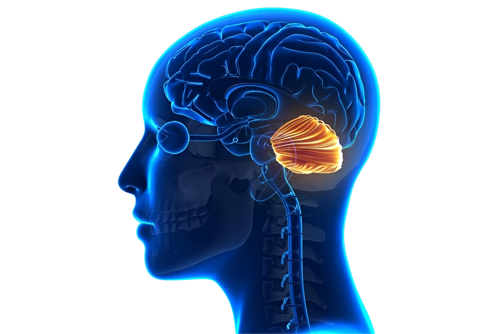
Chiari Malformation, also known as cerebellar herniation, is a structural abnormality in which the balance between the brain and spinal cord is disrupted. It is characterized by the downward displacement of the lower part of the cerebellum, and sometimes the brainstem, from the skull into the spinal canal. Normally, the foramen magnum, an opening at the base of the skull, facilitates the connection between the brain and spinal cord. However, in Chiari Malformation, the cerebellum herniates downward through this opening, obstructing the flow of cerebrospinal fluid (CSF) and causing various neurological problems.
The cerebellum is responsible for balance, movement coordination, and fine motor skills. As a result, pressure exerted by the cerebellum and brainstem on the spinal cord can negatively impact many bodily functions. Symptoms associated with Chiari Malformation can range from headaches to balance issues. Although it is generally considered a congenital disorder, some cases may present during adulthood.
The brain and spinal cord are among the most complex and delicate structures in the body. Cerebellar herniation occurs when these structures develop abnormally due to an anatomical anomaly. Brain structural disorders are often congenital in nature, but external factors such as trauma or tumors can also lead to this condition.
Abnormal development of critical regions such as the cerebellum and brainstem can significantly affect the body’s motor and neurological functions. Cerebellar herniation disrupts the transmission of signals from the brain to the spinal cord, resulting in symptoms such as headaches, muscle weakness, and balance problems. Over time, such structural abnormalities can further strain the nervous system, underscoring the importance of diagnosing and treating Chiari Malformation.
Chiari Malformation is a structural disorder caused by the abnormal descent of the cerebellum and brainstem from the skull into the spinal canal. The condition is classified into four main types, each differing in symptom severity and onset. Type I is the most common, with symptoms that generally begin mildly. Type II is a more severe congenital form, while Types III and IV are rare and associated with more serious health complications.
Type I Chiari Malformation is the most frequently encountered form and is typically diagnosed in childhood or adulthood. It is characterized by the descent of the lower part of the cerebellum (cerebellar tonsils) through the foramen magnum, the opening at the back of the skull, into the spinal canal. In Type I, the brainstem generally remains in its normal position, but cerebellar herniation can exert pressure on the spinal cord, leading to various neurological symptoms.
Type I is typically a congenital disorder but may also develop in some cases due to trauma or excessive cerebrospinal fluid (CSF) drainage. Symptoms may be mild during childhood but can become more pronounced with age. Magnetic Resonance Imaging (MRI) is the most commonly used diagnostic tool. MRI provides detailed and reliable images to assess the degree of cerebellar herniation and its impact on the spinal cord.
Type II Chiari Malformation, also known as Arnold-Chiari Malformation, is a more severe and complex form compared to Type I. In this condition, not only the cerebellum but also the brainstem and the fourth ventricle herniate through the foramen magnum into the spinal canal. Type II is typically congenital and often associated with spinal anomalies such as spina bifida.
Type II is usually a congenital disorder linked to the failure of the spinal cord to close properly during pregnancy (neural tube defect). Prenatal diagnostic methods such as ultrasound can help identify such malformations early. Postnatally, MRI and CT scans evaluate brain and spinal structures to confirm the diagnosis.
Type III Chiari Malformation is extremely rare but also one of the most severe forms. In this type, not only the cerebellum but also the brainstem, and in some cases other parts of the brain, extend from the foramen magnum into the spinal canal. This condition, known as encephalocele, involves the herniation of brain tissue outside the skull, leading to severe neurological dysfunctions.
Type IV Chiari Malformation is characterized by the underdevelopment or absence of the cerebellum. Unlike other types, Type IV is not associated with cerebellar herniation but with incomplete cerebellar development. In Type IV cases, the brain's ability to perform basic motor functions is severely impaired.
The exact causes of these rare types are not fully understood, but they are often linked to congenital anomalies. Prenatal diagnostic methods such as fetal ultrasound and MRI play a critical role in early detection of these abnormalities. After diagnosis, families are often recommended genetic counseling as these anomalies may have hereditary components.
Chiari Malformation is a complex structural disorder that can be caused by congenital or acquired (secondary) factors. It is characterized by the abnormal descent of the cerebellum and brainstem from the skull into the spinal canal. The causes may include genetic predisposition, congenital abnormalities, or factors developing later in adulthood. Below, we will examine the causes and risk factors of Chiari Malformation in detail.
While the exact cause of Chiari Malformation is not fully understood, genetic factors are thought to play a significant role. Several studies have shown that some patients with Chiari Malformation have a family history of similar cases. This suggests that genetic predisposition may trigger the condition. Although genetic transmission is not always direct, it can be linked to other congenital issues such as neural tube defects or spinal anomalies within families.
In conclusion, genetic factors and developmental abnormalities during prenatal growth can create a predisposition to Chiari Malformation. Therefore, individuals with a genetic predisposition should be closely monitored during pregnancy, and necessary precautions should be taken.
Chiari Malformation is generally considered a congenital disorder. During prenatal development, the cerebellum and brainstem may not develop appropriately within the posterior part of the skull. Failure of the neural tube to close properly during pregnancy is one of the primary causes of Chiari Malformation.
These congenital factors are often overlooked during pregnancy but can be detected early through prenatal tests such as ultrasound or MRI. Therefore, regular follow-ups and proper nutrition during pregnancy are of utmost importance.
Acquired Chiari Malformation is a condition that develops in adulthood due to various factors, rather than being present at birth. These malformations can result from trauma, tumors, or issues with cerebrospinal fluid (CSF) drainage, among other external factors.
Acquired Chiari Malformation typically develops suddenly, with symptoms progressing rapidly. Common symptoms in these cases include severe headaches, balance issues, and muscle weakness. Early diagnosis and appropriate treatment approaches are critical for managing acquired cases effectively.
Chiari Malformation presents with a variety of symptoms caused by the abnormal descent of the cerebellum and brainstem into the spinal canal. These symptoms vary depending on the degree of herniation, the pressure on the spinal cord, and the extent to which nerve pathways in the brain are affected. While some patients may experience mild symptoms, others may suffer from severe neurological deficits. Below are the most common symptoms of Chiari Malformation:
Headaches are one of the most frequently reported symptoms among Chiari Malformation patients. These headaches are often triggered or exacerbated by activities such as coughing, sneezing, laughing, or strenuous physical effort. The pain is typically localized at the back of the head and neck region. Such headaches are described as pressure-like and are caused by the disruption of cerebrospinal fluid (CSF) circulation due to the compression of the spinal cord by the cerebellum.
Neck pain is another common symptom. The downward displacement of the cerebellum through the foramen magnum compresses the spinal cord and nerve roots, leading to chronic neck pain. This pain may worsen with movement and significantly impact the patient’s daily life.
Chiari Malformation can cause various neurological symptoms by compressing the spinal cord. This pressure disrupts the normal transmission of nerve signals, leading to neurological dysfunctions in different parts of the body.
Numbness and tingling are common neurological symptoms experienced by patients. These sensations are often felt in the hands, arms, or legs and may affect one or both sides of the body. The compression of the spinal cord by the cerebellum interrupts nerve communication, causing these sensory disturbances.
Balance issues arise due to the cerebellum's role in motor coordination. The cerebellum is responsible for maintaining body balance and fine motor skills. Patients with Chiari Malformation may experience instability while walking, stumbling, and a loss of motor function. These symptoms can severely affect daily functionality and quality of life.
Muscle weakness, particularly in the hands and arms, is another common symptom of Chiari Malformation. The cerebellum and brainstem's disruption of nerve signaling can impair muscle control. As a result, patients may struggle with tasks requiring fine motor skills.
The compression of the spinal cord and disruptions in cerebrospinal fluid (CSF) flow caused by Chiari Malformation can lead to vision and hearing problems. The descent of the cerebellum can impact neural pathways in the brainstem, affecting both vision and hearing functions.
Among the most common vision problems are blurred vision and double vision (diplopia). Due to the cerebellum and brainstem affecting visual pathways, patients may occasionally experience temporary darkening or blurriness in their visual fields. This can make focusing difficult and create challenges in daily activities.
Hearing loss is also observed in some patients with Chiari Malformation. The brainstem’s impact on auditory nerves can reduce hearing capacity or cause tinnitus (ringing in the ears). This issue is often unilateral but may occur bilaterally in some cases.
Chiari Malformation can also cause significant respiratory problems due to the compression of the cerebellum and brainstem into the spinal canal. The brainstem houses centers responsible for controlling respiration, and compression in this area can disrupt respiratory functions.
Sleep apnea is a common respiratory issue in patients with Chiari Malformation. During sleep, patients experience short periods of breathing cessation (apnea), which significantly disrupts sleep quality. Sleep apnea manifests with symptoms such as persistent fatigue, daytime drowsiness, and difficulty concentrating.
Respiratory insufficiency may occur in severe cases of Chiari Malformation. Severe brainstem compression can disrupt the respiratory rhythm, causing shortness of breath. In such cases, respiratory support may be required.
Diagnosing Chiari Malformation involves evaluating symptoms, conducting neurological examinations, and utilizing imaging tests. Patients typically seek medical attention due to headaches, neck pain, or neurological symptoms. These symptoms are assessed through physical and neurological examinations, followed by advanced imaging techniques for a definitive diagnosis.
Patients with Chiari Malformation often present with complaints of headaches, neck pain, balance issues, and neurological symptoms. Initially, the physician conducts a detailed assessment of the patient’s medical history and symptoms. This process is complemented by a physical examination that evaluates the patient’s neurological status.
Key aspects of the physical examination include:
Physical examination can point to specific symptoms of Chiari Malformation, but advanced imaging techniques are required for a definitive diagnosis. If the examination reveals signs of neurological dysfunction, the patient is referred for imaging tests such as MRI or CT.
Magnetic Resonance Imaging (MRI) is the most critical tool for diagnosing Chiari Malformation. MRI provides detailed cross-sectional images of the brain and spinal cord, clearly showing the descent of the cerebellum into the spinal canal. MRI enables a comprehensive evaluation of the severity of cerebellar herniation and associated structural abnormalities.
Computed Tomography (CT) is used to evaluate bone structures and skeletal abnormalities. CT scans assess skull deformities and the positioning of the cerebellum. However, as it does not provide detailed soft tissue images like MRI, it is typically used as a supplementary diagnostic tool.
Cerebrospinal fluid (CSF) analysis is an auxiliary method used in the diagnosis of Chiari Malformation. This is particularly relevant in cases of syringomyelia (fluid-filled cysts in the spinal cord). CSF pressure and composition can provide critical information about the compression exerted by the cerebellum and brainstem on neural pathways. CSF analysis offers insights into how the normal flow of spinal fluid is affected.
Other diagnostic tools include myelography, a specialized radiological test used to visualize spinal cord compression. During myelography, a contrast dye is injected into the spinal canal, enabling detailed imaging of the spinal cord. This test is particularly useful in cases where Chiari Malformation is associated with complications like syringomyelia.
The treatment approach for Chiari Malformation depends on the severity of symptoms, its impact on quality of life, and the extent of cerebellar herniation. Treatment often begins with conservative methods for mild symptoms, but surgical intervention may be necessary for advanced cases. As symptoms can worsen over time, early diagnosis and intervention are crucial. The following section provides a detailed explanation of treatment options and processes.
In cases of Chiari Malformation with mild symptoms, conservative treatment may be preferred over surgical intervention. This approach aims to alleviate symptoms and improve the quality of life for patients. Conservative treatment typically focuses on pain management, physical therapy, and lifestyle modifications.
Conservative treatment methods can be effective for alleviating symptoms; however, patients should be regularly monitored by a physician. If symptoms worsen or neurological damage occurs, surgical treatment should be considered.
Surgical treatment is required when symptoms of Chiari Malformation cannot be controlled with conservative methods or when the progression of the condition leads to neurological damage. Surgical intervention relieves pressure on the cerebellum and brainstem, restoring normal cerebrospinal fluid (CSF) flow.
Posterior fossa decompression surgery is the most common surgical method for treating Chiari Malformation. The primary goal of this procedure is to alleviate the descent of the cerebellum, reduce pressure on neural structures, and restore free CSF flow. The surgery involves the following steps:
Like any surgical procedure, decompression surgery carries certain risks. However, when performed by experienced surgeons, the success rate is high. Potential risks include:
While decompression surgery is the primary method for treating Chiari Malformation, additional surgical procedures may be required in some cases:
Syringomyelia is characterized by the formation of a fluid-filled cyst (syrinx) in the spinal cord. In cases where Chiari Malformation is accompanied by this condition, surgical drainage may be necessary to relieve the pressure caused by fluid accumulation. Treatment of syringomyelia is often performed concurrently with decompression surgery.
If the patient develops hydrocephalus (excessive CSF accumulation in the brain), a shunt may be placed to redirect this fluid to another part of the body. This procedure relieves pressure on the brain and restores fluid balance.
The recovery process following decompression surgery varies depending on the patient's overall health condition and the extent of the surgery. After the procedure, patients are typically monitored in the hospital for several days to ensure close observation.
Chiari Malformation is a condition that requires long-term follow-up and management. Proper treatment and lifestyle adjustments are crucial for improving patients' quality of life. Depending on the severity of symptoms and the type of malformation, individuals living with Chiari Malformation can adapt to their daily lives. This section provides strategies and recommendations for managing life with Chiari Malformation.
Chiari Malformation can affect patients' physical and neurological abilities. However, with appropriate treatment and lifestyle modifications, patients can control their symptoms and enhance their quality of life. Below are some essential strategies for improving quality of life:
Physical activity should be carefully planned for patients with Chiari Malformation. Because cerebellar herniation can cause balance and coordination problems, patients must be cautious during physical activities. However, light and regular exercises can strengthen muscles and reduce pain.
Chiari Malformation is generally a condition that requires long-term monitoring and care. While surgical intervention may control symptoms, long-term follow-up is essential for some patients.

