How is Third Ventriculostomy Performed?
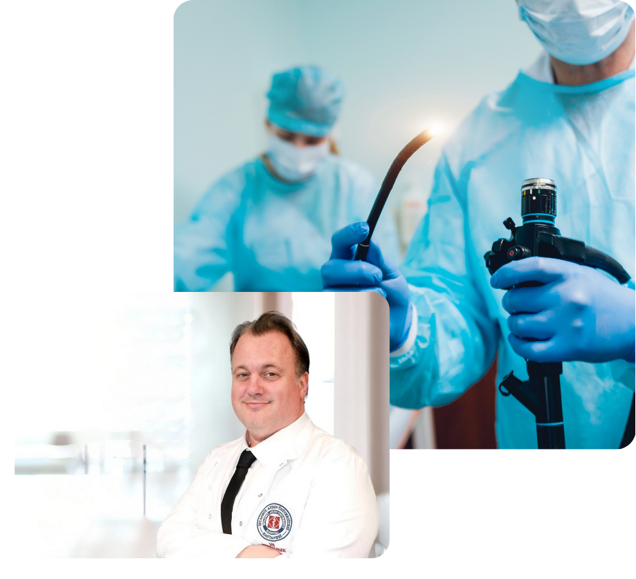
Third ventriculostomy is a surgical procedure performed using an endoscope, which is a thin, flexible tube. During this procedure, the surgeon creates an opening within the brain's ventricles to enable the natural flow of CSF. This allows the fluid to be reabsorbed on the surface of the brain, thereby alleviating the symptoms of hydrocephalus.
- 1. Preparing the Patient: The patient is prepared for surgery under general anesthesia. A small incision is made on the skull, and a hole is drilled in this area.
- 2. Inserting the Endoscope: The surgeon inserts the endoscope through this hole into the brain. The camera at the tip of the endoscope provides a detailed view of the brain's interior, allowing the surgeon to perform the procedure accurately and safely.
- 3. Creating the Third Ventriculostomy: Using the endoscope, the surgeon makes a small opening at the base of the third ventricle. This opening allows the cerebrospinal fluid to flow directly from the brain's ventricles to the base of the brain and then to the surface for absorption.
- 4. Completing the Procedure: Once the opening is made, the endoscope is removed, and the created hole is closed. The surgery typically takes 1-2 hours, after which the patient is transferred to intensive care for monitoring.
ƒ
What Are the Advantages of Endoscopic Third Ventriculostomy?
Third ventriculostomy offers numerous advantages over traditional shunt surgeries, particularly in suitable patients:
- Minimally Invasive: As an endoscopic method, the surgery is minimally invasive. It requires only a small incision, which speeds up recovery and reduces the risk of infection.
- Natural CSF Circulation: This method restores the natural circulation of cerebrospinal fluid, eliminating the need for dependence on a permanent shunt system.
- Lower Risk of Complications: Compared to shunt surgeries, there is a reduced risk of shunt infection, blockage, or malfunction.
- Long-Term Success: ETV can provide long-term success, especially for certain types of hydrocephalus, eliminating the need for frequent shunt replacements.
Who Is It Suitable For?
Endoscopic third ventriculostomy is not suitable for all cases of hydrocephalus. Patients who are most likely to benefit from this procedure typically include:
- Obstructive Hydrocephalus: Cases where CSF flow is blocked due to obstructions in the ventricles.
- Congenital Hydrocephalus: Particularly in conditions like aqueductal stenosis (narrowing of the cerebral aqueduct).
- Patients with Shunt Problems: It may serve as an alternative for patients experiencing recurrent infections or blockages in their shunt systems.
What Are the Risks of Endoscopic Third Ventriculostomy?
Like any surgical procedure, endoscopic third ventriculostomy carries certain risks:
- Bleeding: There is a risk of bleeding in the brain tissue or blood vessels during the procedure.
- Infection: Although it is a minimally invasive method, there is still a risk of postoperative infection.
- Neurological Complications: Neurological impairments may occur due to damage to nerve tissue.
- Failure: In some cases, the created opening may close over time, or it may not adequately support CSF flow, necessitating additional surgical intervention.
Postoperative Recovery Process
The recovery process following endoscopic third ventriculostomy varies depending on the patient’s overall health and the complexity of the surgery. Patients are typically monitored in the hospital for a few days after the procedure. During this period, doctors closely observe the patient to evaluate the success of the surgery.
- Physical Activity: Patients are advised to avoid strenuous physical activities during the first few weeks after surgery. However, light walking is encouraged to promote recovery.
- Imaging Tests: Postoperative MRI or CT scans may be performed to assess the effectiveness of the third ventriculostomy.
- Follow-Up Appointments: Regular follow-up appointments with the neurosurgeon are crucial to ensure continued CSF flow and monitor for the recurrence of hydrocephalus symptoms.
Success Rate of Third Ventriculostomy
Third ventriculostomy has a high success rate, particularly for specific types of hydrocephalus. In patients with obstructive hydrocephalus, such as those with aqueductal stenosis, the success rate can range from 70% to 90%. However, the success of the procedure depends on factors such as the patient’s age, the cause of hydrocephalus, and postoperative care.
Conclusion
Endoscopic third ventriculostomy offers an effective and minimally invasive treatment option for hydrocephalus. With proper patient selection and surgical technique, this method can significantly improve the quality of life for many patients. For individuals diagnosed with hydrocephalus who are seeking an alternative to shunt treatment, this procedure may be a valuable solution. However, the decision for surgery must be carefully evaluated by a specialist, with thorough consideration of patient-specific risks and benefits.

