The Spine and Its Functions
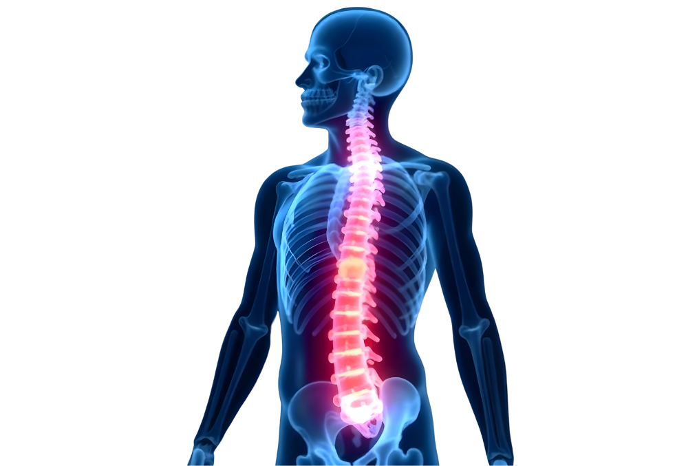
The spine is one of the primary structural components of the human body, providing stability, balance, and flexibility. It also serves as a protective shield for the spinal cord, which is part of the central nervous system. The spine enables the body to stand upright, move flexibly, and supports essential regions such as the head, neck, and torso. Thus, the spine plays a critical role in both mechanical support and neural transmission.
Structure and Sections of the Spine
The spine consists of 33 vertebrae and is divided into five main sections. Each section has specific functions in different regions of the spine:
1. Cervical Region
Comprising 7 vertebrae (C1-C7), this region allows for the movement of the head and is the most flexible part of the spine. The cervical vertebrae enable the neck to move side-to-side and up-and-down. They also support the head and bear its weight. The first two vertebrae, atlas (C1) and axis (C2), are particularly important for neck movement. The atlas supports the skull and enables the "yes" motion, while the axis facilitates the "no" motion.
2. Thoracic Region
Comprising 12 vertebrae (T1-T12), this region connects to the rib bones to form the rib cage, which protects vital organs such as the heart, lungs, and liver. The thoracic spine is less mobile compared to the cervical region and provides more stability. Additionally, the thoracic vertebrae assist in the movement of the ribs during breathing.
3. Lumbar Region
Consisting of 5 vertebrae (L1-L5), this region is the most important for bearing body weight and providing flexibility. The lumbar vertebrae transfer the weight of the upper body to the pelvis and legs. Thus, this region is supported by strong muscles and contributes to movements such as bending forward, backward, and sideways.
4. Sacrum
Made up of 5 fused vertebrae (S1-S5), the sacrum connects to the pelvis and forms a bridge between the upper and lower body. The sacral region constitutes the back wall of the pelvis and transfers body weight to the legs. Additionally, this area serves as a vital passageway for nerve pathways.
5. Coccyx
Composed of 4 fused vertebrae, the coccyx is the lowest part of the spine and is a remnant of the human tail structure. The coccyx is part of the pelvis's support structure and helps the body maintain balance while sitting.
Functions of the Spine
The functions of the spine extend beyond simply supporting and facilitating movement; it also performs several critical life-sustaining roles:
1. Support and Movement
The spine provides solid support to the body and balances body weight during daily activities. Its flexible structure allows for a wide range of movements. The vertebrae in different regions of the spine enable the body to bend forward, backward, sideways, and rotate.
2. Protection
The spine forms a protective canal for the spinal cord. The spinal cord is a critical structure that facilitates the transmission of nerve signals between the brain and the body. Each vertebra encases the spinal cord, shielding it from external impacts. Nerves exit the spinal cord to reach different parts of the body, ensuring neural communication.
3. Flexibility and Resilience
The spine is both a flexible and resilient structure. Its ability to move flexibly enables many essential daily functions. The spinal structure absorbs movement-induced shocks and ensures a balanced distribution of forces. The intervertebral discs act as shock absorbers, protecting the body from impacts during movement.
4. Reflex Control
The spinal cord plays a crucial role in controlling reflex movements. The ability to respond quickly without the brain’s intervention is made possible by the spinal cord. For instance, withdrawing the hand quickly after touching a hot object is a reflex action controlled by the spinal cord before the brain gets involved.
Relationship Between the Spine and Spinal Cord
The spine functions as a protective shield surrounding the spinal cord. The spinal cord transmits nerve signals from the brain to the body and from the body to the brain. This neural transmission ensures the coordination of bodily functions. The relationship between the spinal cord and spine facilitates movement, sensory perception, and reflexes. Nerve roots exit through spaces between the vertebrae and extend to different parts of the body.
The spine is not just a structural support but also a vital connection between the brain and the body. Protecting the spinal cord ensures the proper functioning of neural communication and, therefore, the maintenance of motor abilities.
What are Spinal Tumors?
Spinal tumors are masses characterized by abnormal cell growth in the bones of the spine, the spinal cord, or surrounding tissues. These tumors can exert pressure on the spine or the spinal canal, leading to various health problems. Spinal tumors differ based on their location, cellular structure, and potential for spreading. They can be either benign (non-cancerous) or malignant (cancerous) and can compromise the structural integrity of the spine, causing neurological damage, spinal deformities, and severe pain.
Primary Classification of Spinal Tumors
Spinal tumors are divided into two main categories based on the cells and regions they originate from: primary spinal tumors and secondary (metastatic) spinal tumors. This classification plays a crucial role in understanding where the tumors begin and how they progress.
1. Primary Spinal Tumors
Primary spinal tumors develop directly within the spine or in the tissues surrounding the spinal cord. These types of tumors originate from abnormal cell growth in the bony structures of the spine or the membranes surrounding the spinal cord. Although primary spinal tumors are rare, they can cause significant health issues. They may be benign, but as they grow, they can press on nerve roots and the spinal cord, leading to pain, motor dysfunction, and sensory impairments. Primary spinal tumors may arise from the membranes surrounding the spinal cord, bones, or nerve tissues. Examples include:
- Osteoid Osteoma and Osteoblastoma: Benign tumors originating in the spinal bones but capable of exerting pressure on the spine as they grow.
- Hemangioma: Common benign tumors that develop in the spine, often asymptomatic. However, in rare cases, they can press on nerves, causing pain and movement difficulties.
- Chordoma: A rare malignant spinal tumor that typically occurs in the lower spine, specifically the sacral region, and can grow aggressively.
- Osteosarcoma: A malignant tumor that develops in the spinal bones, spreading rapidly and damaging surrounding tissues.
2. Secondary (Metastatic) Spinal Tumors
Secondary spinal tumors arise when cancer cells from another part of the body spread (metastasize) to the spine. These tumors are the most common spinal tumors and are often caused by cancers originating in the breast, lungs, prostate, kidneys, or thyroid. Cancer cells travel through the bloodstream to the spinal bones, forming metastatic spinal tumors.
- Breast Cancer Metastasis: Breast cancer often spreads to the spinal bones, forming spinal tumors.
- Lung Cancer Metastasis: Lung cancer can metastasize to the spine, causing spinal cord compression and nerve damage.
- Prostate Cancer Metastasis: In prostate cancer patients, the cancer may spread to the spine, leading to back pain, bone fragility, and nerve damage.
Metastatic spinal tumors can compromise spinal integrity, weakening the bone structure and causing fractures. Such fractures may put pressure on the spinal cord, resulting in paralysis, restricted mobility, and neurological dysfunction.
Benign and Malignant Spinal Tumors
Spinal tumors are categorized into two main groups based on their growth rate and ability to spread: benign and malignant tumors.
Benign Spinal Tumors
Benign spinal tumors are slow-growing and typically do not spread to surrounding tissues. However, depending on their size and location, they may compress nerve roots or the spinal cord. This compression can cause symptoms such as pain, muscle weakness, sensory loss, and motor skill impairment. Surgical removal of benign tumors often leads to complete recovery. Nevertheless, some benign tumors may grow over time and lead to serious complications.
- Hemangioma: A benign tumor commonly found in the spinal bones, usually asymptomatic but capable of causing spinal cord compression in rare cases.
- Schwannoma: Benign tumors originating from nerve roots. They grow slowly and can often be completely removed through surgery.
Malignant Spinal Tumors
Malignant spinal tumors are fast-growing and can invade surrounding tissues (metastasize). These tumors can weaken the bone structure, causing spinal fractures and damage to the spinal cord. Malignant spinal tumors may require surgical intervention and are often accompanied by additional treatments such as radiotherapy and chemotherapy.
- Osteosarcoma: A rapidly spreading malignant tumor that originates in the bones, causing damage to both bone and nerve tissues when it develops in the spine.
- Ewing's Sarcoma: A rare malignant bone tumor that usually affects younger individuals and may develop in the spine.
General Effects of Spinal Tumors
Both benign and malignant spinal tumors can affect nerve pathways by exerting pressure on the spinal cord. As a result, patients may experience severe back pain, numbness, tingling sensations, muscle weakness, difficulty walking, and, in advanced cases, paralysis. Depending on the tumor's location, structural deformities in the spine, such as scoliosis, may also occur. Additionally, if the tumor metastasizes, the functionality of other organs may be jeopardized.
Types of Spinal Tumors
Spinal tumors are abnormal cell growths that occur in different regions of the spine and are classified based on their location. The location of spinal tumors plays a significant role in determining their impact on the nervous system and the treatment plan. Generally, spinal tumors are divided into three main categories: extradural, intradural extramedullary, and intradural intramedullary.
1. Extradural Tumors
Extradural tumors develop outside the dura mater (the membrane surrounding the spinal cord) within the bony structures of the spine. These tumors often originate in the spinal bones and may exert direct pressure on the spinal cord. Extradural tumors are the most common site for metastatic tumors. In most cases, they occur due to the spread of cancer from another organ (e.g., breast, prostate, lung, or kidney cancer) to the spine.
- Characteristics:
- Can affect spinal bones, leading to spinal cord compression.
- May cause severe back pain and neurological symptoms due to nerve compression.
- Often metastatic, arising from the spread of cancer from other parts of the body to the spine.
Treatment: Extradural tumors typically require surgical intervention and radiotherapy. If the tumor is metastatic, chemotherapy or targeted therapy may also be applied.
2. Intradural Extramedullary Tumors
Intradural extramedullary tumors are located within the dura mater but outside the spinal cord itself. These tumors can compress the spinal cord and nerve roots, causing neurological damage, although they do not directly harm the spinal cord. The most common tumors in this group include meningiomas and schwannomas.
- Characteristics:
- The tumor forms inside the dura mater surrounding the spinal cord but remains outside the spinal cord itself.
- These tumors are usually benign and grow slowly, but depending on their size, they can cause significant pressure and neurological symptoms.
- Intradural extramedullary tumors can often be successfully treated with surgical intervention.
Common Tumor Types:
- Meningioma: A tumor arising from the meninges (the protective membranes surrounding the spinal cord). Most meningiomas are benign and slow-growing, but they can press on the spinal cord and affect nerve function. They are typically removable through surgery.
- Schwannoma: A benign tumor originating from Schwann cells of the nerve roots. These tumors exert pressure on the nerve tissue and can be surgically removed.
3. Intradural Intramedullary Tumors
Intradural intramedullary tumors develop within the spinal cord, originating from the nerve tissues. These tumors directly damage the nerve cells within the spinal cord, potentially leading to severe neurological deficits. This category includes some of the most dangerous types of spinal tumors, as they directly affect spinal cord tissue and disrupt nerve pathways.
- Characteristics:
- Tumors located within the spinal cord that directly compress nerve cells.
- These tumors can be either benign or malignant, but in both cases, they create pressure on the spinal cord and cause severe neurological issues.
- The delicate nature of the nerve tissue makes treatment challenging.
Common Tumor Types:
- Astrocytoma: A tumor that arises from nerve cells within the spinal cord. Astrocytomas can be benign or malignant. They create pressure on spinal nerve tissues, potentially leading to movement and sensory impairments.
- Ependymoma: A tumor developing in the spinal canal where cerebrospinal fluid flows. Ependymomas are usually benign but can press on the spinal cord as they grow.
- Hemangioblastoma: A rare tumor originating from the blood vessels within the spinal cord. Often associated with Von Hippel-Lindau disease, hemangioblastomas require surgical intervention.
Common Types of Spinal Tumors
Spinal tumors vary in location and tissue characteristics. Below are common types of spinal tumors and their features:
- Meningioma: A benign tumor arising from the meninges (membranes surrounding the spinal cord). These tumors typically grow slowly and can be effectively treated with early diagnosis and surgical removal.
- Schwannoma: A benign tumor developing from Schwann cells, which form the nerve sheaths. Schwannomas can exert pressure on the nerve roots emerging from the spinal cord and are often surgically removable.
- Astrocytoma: A tumor arising from nerve cells within the spinal cord. Astrocytomas can be benign or malignant. Treatment typically involves surgery, radiation therapy, or chemotherapy.
- Ependymoma: A benign tumor arising from cells lining the cerebrospinal fluid pathways within the spinal canal. As these tumors grow, they can compress the spinal cord, causing neurological issues.
- Chordoma: A rare, malignant tumor that forms in the lower part of the spine, especially the sacrum. Chordomas grow slowly but can damage surrounding tissues and bone structures. These tumors are challenging to treat and often require surgery and radiotherapy.
Symptoms of Spinal Tumors
Symptoms of spinal tumors vary significantly depending on the size, location, and pressure they exert on the nerve tissues. In the early stages, symptoms may be mild and gradually worsen over time. Spinal tumors typically press on the spinal cord, nerve roots, or spinal bones, leading to both localized and neurological symptoms.
- Persistent Back Pain:
Back pain is the most common symptom of spinal tumors. This pain results from the tumor pressing on the spinal bones or spinal cord. As the tumor grows and applies more pressure on the nerve roots, the intensity of the pain increases. Tumor-related back pain often has the following characteristics:
- Worsens at night: Tumor-related pain is often more intense at night or during periods of rest.
- Chronic: The pain is persistent, does not subside, and worsens over time.
- Not activity-dependent: Unlike other types of back pain, tumor-related pain is not linked to physical activity and is often felt even during rest.
- Numbness and Tingling:
Tumors can disrupt the function of the spinal cord or nerve roots, leading to loss of sensation in specific areas of the body. Numbness and tingling symptoms typically occur in the following regions depending on the tumor's location:
- Numbness in the arms and legs: When nerve pathways are affected, patients may experience tingling and loss of sensation in the arms, legs, hands, and feet.
- Localized sensory loss: Depending on the tumor's location, spinal cord tumors can cause sensory loss in specific regions of the body, such as the chest area or below the waist.
- Muscle Weakness:
Spinal tumors can impair nerves, leading to muscle weakness. The pressure exerted by the tumor on the spinal cord or nerve roots can hinder the ability of nerves to send effective signals to muscles, resulting in muscle weakness. Symptoms of muscle weakness include:
- Difficulty walking: Weak muscles make walking challenging and may cause instability.
- Weakness in the hands and legs: Loss of strength in the arms, legs, and hands may occur, complicating daily tasks.
- Balance issues: Muscle weakness can cause instability and frequent falls.
- Walking Difficulties and Balance Problems:
Spinal tumors may interfere with the proper functioning of nerves along the spinal cord, leading to walking and balance issues. Tumors in the lumbar or thoracic spine may affect the lower body, resulting in the following symptoms:
- Lack of coordination: Difficulty maintaining coordination during walking may cause a slow and unsteady gait.
- Frequent falls: Balance issues increase the risk of falling.
- Advanced Symptoms:
If untreated or if the tumor continues to grow, more severe neurological symptoms can develop:
- Paralysis: Spinal tumors may compress the spinal cord, disrupting nerve signal transmission. This can lead to partial or complete paralysis depending on the tumor's location:
- Lower body paralysis (paraplegia): Tumors in the lumbar region may paralyze the legs.
- Complete body paralysis (quadriplegia): Tumors in the cervical spine can affect all four limbs, including the arms and legs.
- Loss of Bladder and Bowel Control: Tumors in the lower spine may press on nerves controlling bladder and bowel functions, causing incontinence and bowel dysfunction:
- Bladder control issues: Patients may experience difficulty managing urination, leading to incontinence.
- Bowel control issues: Similar issues may arise with bowel movements, causing irregularities and loss of control.
- Spinal Deformities: Large tumors can weaken the structural integrity of the spine, leading to scoliosis or kyphosis. These deformities alter the natural curvature of the spine and affect posture:
- Scoliosis: The spine may curve to one side due to structural changes caused by the tumor.
- Kyphosis: Tumors in the thoracic region can cause the spine to bend forward, leading to a hunched posture.
Causes and Risk Factors of Spinal Tumors
The exact causes of spinal tumors remain unclear, but genetic factors, environmental exposures, and other medical conditions may play a significant role in their development. Spinal tumors arise from abnormal cell growth in the spinal bones or surrounding tissues and can be categorized as primary (originating in the spine or spinal cord) or secondary (metastatic tumors spread from other organs). The key causes and risk factors contributing to spinal tumors include:
- Genetic Factors:
Certain genetic disorders and inherited conditions significantly increase the risk of spinal tumors. These conditions often show a higher prevalence of spinal and spinal cord tumors:
- Von Hippel-Lindau Syndrome (VHL): VHL is a rare genetic disorder that causes tumors and cysts to form throughout the body. It particularly predisposes individuals to tumors in the spine and brain. People with VHL have a high incidence of spinal tumors.
- Neurofibromatosis Type 2 (NF2): NF2 is a genetic disorder characterized by the development of tumors in the nervous system. It commonly leads to tumors around the spine, such as schwannomas.
- Li-Fraumeni Syndrome: This genetic disorder increases the risk of various cancers, including spinal tumors. Caused by mutations in the TP53 gene, it heightens the likelihood of tumor development in multiple parts of the body.
- Radiation Exposure:
Radiation can damage cellular DNA and trigger mutations, leading to tumor development. High-dose radiation exposure is a significant environmental factor that increases the risk of spinal tumors:
- Previous Radiation Therapy: Radiation treatments for cancer can cause long-term damage to spinal or spinal cord tissues. Radiation therapy is a known risk factor for tumors developing in sensitive tissues like bone marrow or the spinal cord.
- Environmental Radiation Exposure: Individuals exposed to high doses of radiation, such as those working near nuclear plants or in environments with high radiation levels, are at an increased risk of developing spinal tumors due to DNA damage.
- Metastatic Tumors:
Metastatic tumors form when cancer cells from other parts of the body spread to the spine. The spine is particularly vulnerable to metastatic cancer from other regions, and various cancer types can spread to the spine:
- Breast Cancer: Breast cancer frequently spreads to the spine in advanced stages, causing spinal tumors through bone metastasis, which can compromise the structural integrity of the spine.
- Prostate Cancer: Prostate cancer often metastasizes to the spine, exerting pressure on the spinal bones and causing pain, bone weakening, and neurological complications.
- Lung Cancer: Lung cancer is a common cause of metastatic spinal tumors. Cancer cells from the lungs can travel to the spine and form new tumors.
- Kidney Cancer: Kidney cancers can metastasize to the spine, damaging the spinal bones. These metastases often cause back pain, muscle weakness, and issues with spinal stability.
- Other Cancers: Cancers such as melanoma, lymphoma, and gastrointestinal cancers can also metastasize to the spine. These metastatic tumors can harm the spinal bones and nerve tissues, resulting in a range of symptoms.
- Immune System Disorders:
The immune system defends the body against abnormal cell growth. However, when the immune system is compromised, cancerous cells are more likely to grow and spread:
- Immune Deficiency: A weakened immune system reduces the body's ability to combat cancerous cells, increasing the risk of spinal tumors. Conditions such as HIV/AIDS can predispose individuals to cancer development.
- Autoimmune Diseases: Certain autoimmune diseases, where the immune system mistakenly attacks its own tissues, can lead to spinal tumors. These conditions may disrupt normal immune regulation.
- Other Risk Factors:
Additional risk factors for the development of spinal tumors include:
- Age: Advanced age is a risk factor for spinal tumors. As people age, the likelihood of genetic mutations in cells increases, which can lead to the formation of spinal tumors.
- Exposure to Chemicals: Prolonged exposure to chemical substances or carcinogens in the workplace increases the risk of spinal tumors.
- Family History: Individuals with a family history of spinal tumors may have a higher risk of developing tumors due to hereditary factors.
How Are Spinal Tumors Diagnosed?
The diagnosis of spinal tumors depends on the patient’s symptoms, the location, and the size of the tumor. Accurate diagnosis is critical for understanding the nature and spread of the tumor and for developing an appropriate treatment plan. The diagnostic process includes imaging techniques, tests to assess nerve function, and invasive methods such as biopsy. The main diagnostic methods include:
- MRI (Magnetic Resonance Imaging):
MRI is the most commonly used and sensitive imaging method for diagnosing spinal tumors. It allows for detailed examination of the spine and spinal cord, providing critical information about the size, location, and effects of the tumor on surrounding tissues.
- Detailed Imaging of the Spinal Cord and Nerve Tissues: MRI is especially effective in visualizing soft tissue structures and provides detailed images of the spinal cord and nerve roots. It reveals the tumor’s pressure on nerves, proximity to the spinal cord, and potential spread.
- Tumor Characterization: MRI images help evaluate the structural features of the tumor and determine whether it is benign or malignant.
- Use of Contrast Agents: During MRI scans, contrast agents may be used to enhance the visibility of the tumor. This makes the tumor tissue more distinguishable and aids in understanding the effects of metastatic tumors on the spine.
- CT (Computed Tomography):
Computed tomography (CT) is a widely used imaging technique for examining the bony structures of the spine. CT scans are particularly useful for evaluating the impact of tumors on bone structures and assessing potential spread to spinal bones.
- Examination of Spinal Bones: CT provides detailed imaging of bone tissue, helping to identify the pressure, deformation, or destruction caused by the tumor on spinal bones. This is especially important in cases of metastatic tumors, which tend to damage bone tissue.
- Evaluation of Structural Damage in the Spine: CT can detect weakening of bones, fractures, or structural damage that threatens spinal stability. This information is crucial for planning surgical interventions.
- Contrast-Enhanced CT: The use of contrast agents highlights the differences between tumor tissue and healthy bone tissue, providing more detailed information about the size and extent of the tumor.
- Biopsy:
A biopsy is an invasive procedure used to analyze samples of tumor tissue to determine its type, characteristics, and malignancy. Biopsies are typically performed before surgery or in suspicious cases and can be conducted through the following methods:
- Fine Needle Biopsy: A minimally invasive method where a small tissue sample is taken from the tumor. It is usually performed under local anesthesia and results can be obtained quickly.
- Open Biopsy: If a fine needle biopsy is insufficient or a larger tissue sample is needed, an open biopsy is performed. This involves a surgical procedure to collect a larger sample from the tumor.
- Pathological Examination: The biopsy sample is examined under a microscope to assess cell structure, growth rate, and whether the tumor is benign or malignant. Biopsy results play a critical role in shaping the treatment plan.
- Neurological Tests:
Neurological tests are also used during the diagnostic process to evaluate the effects of spinal tumors on the nervous system. Understanding the extent of nerve function impairment helps determine the severity of the tumor and its treatment requirements.
- EMG (Electromyography): EMG measures nerve and muscle function, assessing the pressure exerted by the tumor on nerve roots. By analyzing the electrical activity of muscles, it reveals the extent of nerve damage.
- SEP (Somatosensory Evoked Potentials): SEP measures sensory functions of nerve pathways, identifying disruptions caused by the tumor. This test helps determine the extent of nerve damage and whether nerve functions can be restored.
- Other Imaging Techniques:
Other imaging techniques include:
- PET-CT (Positron Emission Tomography-Computed Tomography): PET-CT measures the metabolic activity of cancer cells and is used to determine whether tumors are metastatic. This imaging technique is essential for evaluating the potential spread of spinal tumors to other parts of the body. PET-CT identifies the location and activity of malignant tumors, optimizing the treatment plan.
- The Importance of Early Diagnosis:
Early diagnosis of spinal tumors directly impacts treatment outcomes. Detecting tumors in their early stages allows for more successful surgical or other therapeutic interventions. Intervening when the pressure on nerve tissues is minimal increases the chances of preserving neurological functions. Early diagnosis also reduces the likelihood of metastasis and significantly improves patients' quality of life.
Treatment Methods for Spinal Tumors
Treatment methods for spinal tumors are determined based on the type, size, location of the tumor, the patient’s overall health condition, and the impact of the tumor on nerves, the spinal cord, and surrounding tissues. Treatment typically involves a combination of surgical intervention, radiotherapy, chemotherapy, and supportive therapies. The most appropriate treatment approach is individualized for each patient by a multidisciplinary team, with a patient-centered approach adopted throughout the treatment process.
- Surgical Intervention:
Surgical intervention is a commonly used treatment method for spinal tumors, especially for large tumors causing nerve compression. The goal of surgery is to remove the tumor and relieve pressure on the spinal cord. Surgery is often necessary to restore spinal stability and preserve nerve functions. Situations where surgery is preferred include:
- Large Tumors: Large tumors that cause neurological symptoms by compressing the spinal cord or nerves can be surgically removed.
- Preservation of Nerve Functions: Surgery helps alleviate pressure on nerve roots, aiding in the preservation of neurological functions.
- Restoration of Stability: If the tumor has caused significant damage to the spinal structure, stabilization of the spine may be required. Metal rods, plates, or screws are used to prevent spinal collapse.
Two main techniques are used in surgical interventions: microsurgery and minimally invasive surgery:
- Microsurgical Techniques: Enables surgeons to remove the tumor with high precision under a microscope. This technique is preferred for tumors close to nerve roots. Microsurgery minimizes damage to surrounding tissues and carries a lower risk of complications.
- Minimally Invasive Surgery: Performed through small incisions, this method speeds up recovery time and reduces the risk of complications. Minimally invasive surgery is particularly beneficial for benign tumors, allowing for quicker recovery.
Surgical intervention is often combined with radiotherapy and chemotherapy for metastatic or malignant tumors. Postoperative physical therapy and rehabilitation play a vital role in recovering nerve functions.
- Radiotherapy:
Radiotherapy is used when surgical intervention is not possible or to eliminate residual tumor cells after surgery. Radiotherapy targets tumor cells to stop their growth or shrink them. Radiotherapy is applied in the following situations:
- Post-Surgery: Radiotherapy is used if the tumor cannot be completely removed or if there is a high risk of recurrence.
- When Surgery Is Not Feasible: Radiotherapy is preferred if the tumor's location makes surgery risky or if the patient’s health condition is not suitable for surgery.
Stereotactic Radiosurgery (CyberKnife): This modern version of radiotherapy focuses high-dose radiation precisely on the tumor:
- CyberKnife Radiosurgery: This technique delivers high-dose radiation directly to the tumor without damaging surrounding healthy tissues. CyberKnife is particularly effective for spinal tumors, ensuring the preservation of surrounding tissues and enhancing treatment efficacy.
- Chemotherapy:
Chemotherapy is used to treat malignant and metastatic spinal tumors. Chemotherapy aims to kill or inhibit the growth of tumor cells using drugs. Chemotherapy is typically applied in the following cases:
- Metastatic Tumors: Chemotherapy helps control tumors that have spread to the spine from other organs.
- Combined Therapy: Chemotherapy is used alongside surgery and radiotherapy to stop tumor growth and reduce the risk of recurrence.
- Treatment of Malignant Tumors: For malignant tumors within spinal bones, chemotherapy is employed to halt their spread.
- Supportive Therapy:
Treatment of spinal tumors aims not only to remove the tumor but also to improve patients’ quality of life and alleviate symptoms through supportive therapies. Supportive treatments include:
- Pain Management: Spinal tumors can cause severe pain. Effective pain management significantly enhances patients’ quality of life. Pain management methods include medication (analgesics), nerve blocks, and epidural steroid injections.
- Spinal Stabilization: Spinal tumors can weaken the spinal structure, requiring stabilization. Surgical intervention stabilizes the spine to prevent structural collapse, often using metal implants.
- Physical Therapy and Rehabilitation: Post-surgical physical therapy programs are implemented to restore nerve functions and improve muscle strength. Rehabilitation helps patients return to daily life more quickly.
- Psychological Support: Treating spinal tumors is a lengthy and challenging process. Psychological support is often necessary for patients. Psychological counseling and group therapies can help patients navigate the treatment process more effectively.
- Multidisciplinary Approach:
A multidisciplinary approach is essential in the treatment of spinal tumors. Oncologists, surgeons, radiologists, neurologists, physical therapy specialists, and psychologists work together to create the most appropriate treatment plan for the patient. This teamwork ensures the development of the most effective treatment strategy, taking into account the tumor type, location, and the patient's overall condition.
Since every patient's condition is different, creating personalized treatment plans through a multidisciplinary approach plays a critical role in improving treatment success and enhancing the patient's quality of life.
Postoperative Recovery Process and Follow-Up
The recovery process after spinal tumor surgery can vary significantly depending on the type of surgery, tumor location, and the patient's overall health. Physical therapy, rehabilitation, and regular medical check-ups play a critical role during this period. Managing the recovery process helps patients regain spinal function, reduce pain, and improve quality of life.
- Postoperative Recovery Process:
After surgery, patients are typically monitored in the hospital for a few days to a few weeks. This period varies based on the scope of the surgery, the patient's preoperative health, and the damage caused by the tumor to nerve tissues. Key points to consider during the recovery process include:
- Early Mobilization: Post-surgery, it is recommended that patients get up and start light movements as soon as possible. This increases blood circulation, preventing clot formation and muscle loss. In minimally invasive surgeries, patients recover faster and can start walking within a few days.
- Physical Therapy and Rehabilitation: Physical therapy and rehabilitation programs after spinal tumor surgery are vital in helping patients regain nerve functions. These programs are designed to support spinal stabilization, restore muscle strength, and improve balance and coordination. Physical therapy begins immediately after surgery and may last weeks or months, depending on the patient's condition.
- Spinal Stabilization: Stabilization methods such as metal rods, screws, or plates used during surgery help strengthen the spine. However, patients should support spinal movements with specific exercises and avoid heavy activities that may strain the spine during this period.
- Potential Complications:
Some complications may occur during the postoperative period. Detecting and managing these complications early helps patients go through the recovery process more smoothly. Possible complications include:
- Nerve Damage: Spinal tumors are located close to nerve tissues, making nerve damage a risk during surgery. Postoperative nerve damage can lead to neurological issues such as muscle weakness, sensory loss, or paralysis. In such cases, physiotherapy and rehabilitation programs can help recover some nerve functions.
- Infection: As with any surgical procedure, there is a risk of infection post-surgery. Therefore, maintaining the cleanliness of the surgical site, performing regular dressings, and using prescribed antibiotics are essential. Signs of infection include swelling, redness, fever, and pain at the surgical site.
- Tumor Recurrence: Spinal tumors may recur after surgical intervention in some cases. Regular follow-ups and imaging techniques such as MRI or CT scans are necessary to monitor the tumor's status. For patients with a high risk of recurrence, postoperative radiotherapy or chemotherapy may be planned.
- Postoperative Pain: Post-surgical pain is common and can usually be managed with painkillers. However, prolonged or worsening pain may require further evaluation. Pain management is an essential part of recovery and may include methods such as epidural injections or nerve blocks.
- Follow-Up and Regular Check-Ups:
Regular follow-ups and long-term monitoring are vital for patients after spinal tumor surgery. During medical check-ups, the patient’s overall health, nerve functions, and spinal stability are evaluated. These follow-ups typically include:
- Imaging Tests: Techniques such as MRI, CT, or PET-CT are used to monitor the tumor’s status. Postoperative imaging ensures the tumor has been completely removed and assesses the minimal risk of regrowth.
- Neurological Evaluation: The extent of nerve function recovery is assessed through neurological tests. Reflexes, muscle strength, balance, and sensory tests are part of these evaluations.
- Hormonal and Metabolic Tests: Spinal tumors affecting the spinal cord or nerve tissues can disrupt hormonal balance in the body. Physicians regularly monitor hormone levels and make necessary adjustments to treatment.
- Lifestyle Changes and Recovery Period:
After surgery, patients may need to make certain lifestyle changes. To accelerate recovery and maintain spinal health, attention should be paid to the following:
- Exercise: Exercises within physical therapy programs enhance muscle strength and flexibility. However, heavy lifting, sudden movements, and strenuous sports activities should be avoided.
- Balanced Diet: Adequate intake of protein, vitamins, and minerals supports the recovery process. Increased consumption of calcium and vitamin D is particularly important for bone and muscle health.
- Stress Management: Stress can negatively affect the recovery process after surgery. Techniques such as yoga and meditation can be used for stress management.
- Long-Term Care and Psychological Support:
Long-term care after spinal tumor surgery is necessary to improve patients’ quality of life. While physical therapy and rehabilitation programs improve nerve functions, psychological support helps maintain morale and motivation.
- Psychological Support: Spinal tumor surgery may lead to emotional challenges such as anxiety and depression. These conditions can be managed during the postoperative period with the help of a psychologist or psychiatrist. Psychological support contributes to better adaptation to the treatment process and a healthier recovery period.
- Long-Term Follow-Up: Patients must continue regular medical check-ups after surgery. The risk of tumor recurrence, general health, and nerve functions are periodically assessed. This follow-up enables early detection and treatment of tumor recurrence.
Conclusion
The recovery process after spinal tumor surgery requires a multidisciplinary approach. Physical therapy, neurological rehabilitation, and regular follow-ups aid in regaining nerve functions and improving patients’ quality of life. Preventing potential complications during the postoperative period and ensuring long-term follow-up minimizes the risk of tumor recurrence.