Logo


Icon

Call Now

0540 930 20 20

Icon

Email

info@gucluhanguclu.com

Icon

Address

Beşyol, Florya, Akasya St. No:4 Apt:1, 34295 Küçükçekmece/Istanbul

FOLLOW US

Stay updated with the latest news about Assoc. Prof. Dr. Güçlühan Güçlü

Neuro-Oncology

Spinal Cord Tumor

Comprehensive information about spinal cord tumors and treatment options.

Bel Fıtığı Tedavisi

Spinal Cord Tumor

What is a Spinal Cord Tumor?

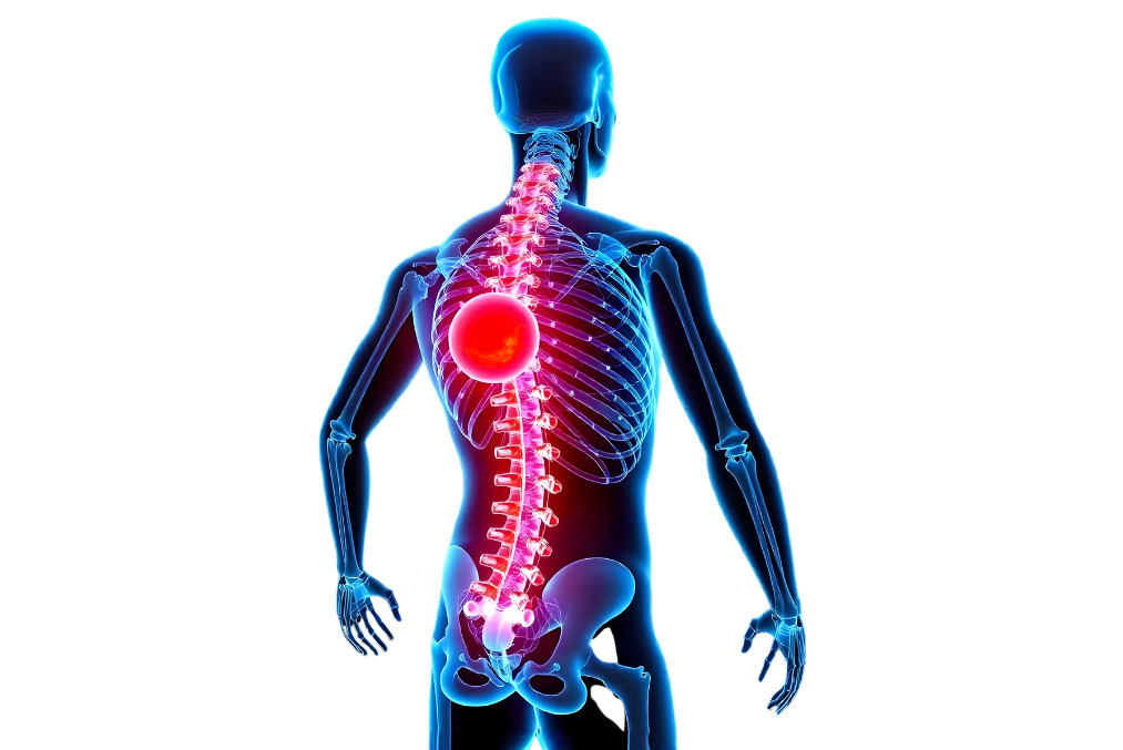
A spinal cord tumor is a condition characterized by abnormal cell growth in the spinal cord or its membranes (meninges). The spinal cord is a structure composed of nerve cells that transmits signals between the brain and the rest of the body. Spinal cord tumors can compress nerve fibers, leading to impairments in neurological function. These tumors can originate directly from spinal cord cells or spread from the surrounding bones, tissues, or other regions.

Omurilik Tümörleri

The Spinal Cord and Its Function

The spinal cord plays a vital role as part of the central nervous system. It facilitates signal transmission between the brain and the body, regulating various bodily functions. The spinal cord begins at the brainstem and extends downward through the spine. Measuring approximately 45 cm in length, its primary functions include transmitting motor and sensory signals, controlling reflexes, and coordinating muscle movements.

Signal Transmission and Reflex Control

The spinal cord enables the two-way flow of signals between the brain and the body. Motor signals are generated by the brain and transmitted to the muscles through the spinal cord, enabling body movement. Simple motor actions, such as raising your hand or running, are achieved this way. Sensory signals, on the other hand, are sent from the body to the brain. Information such as touch, temperature, pressure, and pain from various areas like the skin, muscles, joints, and organs reaches the brain via the spinal cord. The brain processes this information to enable the body to respond appropriately.

The spinal cord is also the center of reflexes. Reflexes are managed quickly by the spinal cord without requiring conscious input from the brain. For example, when you touch a hot surface, the spinal cord rapidly sends a motor signal to withdraw your hand. This reflex action is executed without the signal traveling to and from the brain, allowing for faster response times and immediate protection of the body.

Regional Functions of the Spinal Cord

The spinal cord serves different body regions at various levels. It consists of four main sections:

  1. Cervical (neck) spinal cord: This section starts from the neck and transmits nerve signals to the upper arms and hands. It also plays a vital role in controlling the diaphragm, which is essential for breathing.
  2. Thoracic (chest) spinal cord: This region is associated with the torso, abdominal, and back muscles. It is crucial for maintaining posture and balance.
  3. Lumbar (lower back) spinal cord: The nerves in this section control the lower extremities, such as the legs and feet. Activities like walking, running, or controlling leg muscles rely on signals from the lumbar region.
  4. Sacral (pelvic) spinal cord: The sacral region governs the functions of pelvic organs, including urination, bowel movements, and sexual functions.

This regional structure highlights the critical role of the spinal cord's nerve network and how it carries signals to every part of the body.

The Relationship Between the Spine and the Spinal Cord

The spine consists of rigid bone structures that protect the spinal cord. Vertebrae function as a protective shield for the spinal cord, safeguarding it from mechanical damage, impact, or injury. Each vertebra contains a nerve branch from the spinal cord; these branches exit the vertebrae to serve specific areas of the body. For example, nerves from cervical vertebrae in the neck reach the upper extremities, while those from lumbar vertebrae in the lower back control the lower extremities.

Discs located between vertebrae balance the pressure on the spine, allowing the spinal cord to function smoothly. These discs enhance spinal flexibility and mobility. Issues like disc degeneration or herniation can exert pressure on the spinal cord, disrupting nerve transmission and impairing spinal cord functions.

The Spinal Cord as a Neural Highway

The spinal cord can be described as a "neural highway" connecting the brain and the body. The transmission of sensory information to the brain and motor commands from the brain to the body occurs along this highway. The nerve pathways in the spinal cord are categorized into two main groups:

  • Dorsal nerve pathways (posterior roots): Carry sensory signals.
  • Ventral nerve pathways (anterior roots): Transmit motor signals.

This neural highway system allows the brain to maintain full control over the body. However, any interruption at various levels of the spinal cord can result in significant motor and sensory dysfunctions. For instance, a spinal cord injury at the neck level may cause paralysis and sensory loss in most of the body, whereas an injury at the lower back typically results in impairments limited to the legs and pelvic functions.

The Vital Role of Reflexes

Reflexes are quick, automatic responses essential for survival. The spinal cord manages these responses through a neural pathway called the reflex arc. Reflexes occur without the need for signal transmission to the brain. For example, the sudden upward jerk of the foot when the knee is tapped is a reflex response controlled by the spinal cord. These reflexes allow the body to react quickly to potential dangers and protect itself.

What is a Spinal Cord Tumor?

A spinal cord tumor is an abnormal, uncontrolled growth of cells within the spinal cord tissue or surrounding areas. These tumors can damage the spinal cord's delicate structure, impair nerve function, and disrupt nerve signal transmission, leading to various neurological symptoms. The severity of symptoms caused by spinal cord tumors depends on their size, location, and nature.

Types of Spinal Cord Tumors

Spinal cord tumors are classified based on their location, origin, and cellular structure. Broadly, there are two main types of tumors:

  1. Primary Spinal Cord Tumors: These tumors originate within the spinal cord or the tissues surrounding it. Tumors occurring in or near the spinal cord disrupt its natural structure and directly affect nerve functions. Primary tumors are often benign but can cause severe neurological damage if they grow or press on nerves.
  2. Metastatic Spinal Cord Tumors: Metastatic tumors develop when cancer from another part of the body spreads to the spinal cord. Cancers originating from organs such as the lungs, breasts, prostate, and kidneys can metastasize to the spinal cord. These tumors are often malignant and rapidly affect spinal cord functions, leading to severe symptoms.

Benign and Malignant Spinal Cord Tumors

Spinal cord tumors are also classified as benign (non-cancerous) or malignant (cancerous) based on their cellular characteristics:

  1. Benign Tumors: These tumors typically grow slowly and do not spread to surrounding tissues. However, despite being benign, they can cause significant problems by pressing on nerves when growing in the confined and critical space of the spinal cord. They can disrupt nerve signal transmission and lead to neurological deficits. Most benign tumors can be surgically removed, often resulting in full recovery.

    Common Types of Benign Tumors:

    • Meningioma: Develops in the membrane layer surrounding the spinal cord and typically grows slowly. It is often benign and can be completely removed surgically.
    • Schwannoma: Benign tumors originating from nerve roots. They develop from the cells of the nerve sheath and can be treated with surgical intervention.
    • Hemangioblastoma: Benign tumors arising from the blood vessels of the spinal cord.
  2. Malignant Tumors: Malignant tumors are aggressive, rapidly growing, and tend to spread to surrounding tissues. These tumors can damage nerve tissue and lead to severe neurological disorders. If left untreated, they can cause permanent damage to the spinal cord, nerves, and spine.

    Common Types of Malignant Tumors:

    • Astrocytoma: A malignant tumor growing within the spinal cord, often originating from nerve cells. It frequently requires surgical intervention and radiotherapy.
    • Ependymoma: Develops from ependymal cells within the spinal cord. It can be benign or malignant but may damage surrounding tissues as it grows.
    • Metastatic Tumors: Malignant tumors that spread to the spinal cord from other organs. If untreated, they can quickly cause loss of neurological function.

Differences Between Primary and Metastatic Tumors

Primary tumors arise from the abnormal growth of cells within the spinal cord or its membranes. Since these tumors originate directly from spinal cord tissue, they spread more slowly to surrounding tissues. While they can be benign, primary tumors may press on nerves as they grow, impairing motor and sensory nerve functions.

Metastatic tumors, on the other hand, result from the spread of cancerous cells from other parts of the body to the spinal cord. These tumors originate from primary cancer sources such as the lungs, breasts, or kidneys and form new tumor sites in the spinal cord. Metastatic tumors are typically malignant and spread quickly. If untreated, they can cause nerve damage, motor function loss, paralysis, and life-threatening complications.

The Impact of Spinal Cord Tumors on Neurological Functions

Spinal cord tumors disrupt nerve signal transmission, leading to impairments in normal bodily functions. As they press on nerves, patients may experience symptoms such as pain, muscle weakness, sensory loss, and balance issues. As the tumor grows, these symptoms can worsen, potentially resulting in paralysis, loss of bladder control, bowel problems, and sexual dysfunctions.

The Importance of Treating Spinal Cord Tumors

Prompt diagnosis and treatment of spinal cord tumors are critical to preventing nerve damage. Since the spinal cord is located in a confined space, tumor growth can block nerve pathways and affect vital functions. Untreated tumors can severely reduce a patient’s quality of life and cause permanent neurological damage. Early diagnosis and treatment of spinal cord tumors minimize functional losses and help patients return to their normal lives.

Symptoms of Spinal Cord Tumors

Spinal cord tumors can present a wide range of symptoms depending on their location, size, and the affected nerve pathways. These symptoms are typically caused by pressure on the spinal cord and vary according to the affected area of the spinal cord. These tumors, which can have significant impacts on nerve transmission and body functions, manifest through various symptoms in both early and advanced stages.

  1. Persistent Back Pain:

    One of the most common symptoms of spinal cord tumors is back pain. This pain is typically localized to the segment of the spinal cord where the tumor is situated and worsens as the tumor grows. Initially, the pain may be mild and intermittent, but over time, it can become constant and unbearable. Back pain caused by spinal cord tumors can persist even during rest and may become more pronounced at night. This pain results from the tumor growing and exerting pressure on the spinal cord and nerves.

    • Localized pain: Pain concentrated in the area of the tumor, often described as deep and continuous.
    • Radicular pain: Tumors pressing on nerve roots can cause pain that radiates from the back to the legs, arms, or other parts of the body. This pain is often sharp and burning in nature.
  2. Numbness and Tingling:

    Spinal cord tumors can exert pressure on nerve pathways, causing sensory symptoms such as numbness and tingling in specific areas of the body. These symptoms are typically a result of the tumor blocking affected nerve pathways. Numbness, tingling, or a burning sensation may occur in the upper or lower parts of the body. These symptoms often worsen as pressure on the spinal cord increases and can interfere with nerve function.

    • Numbness in the arms and legs: As the tumor grows, it obstructs nerve transmission, leading to numbness, burning, or tingling in the arms, legs, or other parts of the body.
    • Loss of skin sensation: In some cases, sensitivity in the skin may decrease, impairing the ability to perceive sensations such as cold, heat, or touch.
  3. Muscle Weakness:

    Spinal cord tumors pressing on nerve roots can affect motor nerve pathways, causing muscle weakness and loss of strength. Muscle weakness usually occurs in specific parts of the body, depending on the affected section of the spinal cord. For example, a tumor in the lower spine may cause weakness in the leg muscles, while a tumor in the upper spinal cord may result in arm weakness. In advanced cases, muscle atrophy and significant reductions in mobility may occur.

    • Weakness in the arms and legs: Muscle weakness, particularly in the legs, may lead to difficulties with walking.
    • Loss of fine motor skills: Weakness in the hands may cause difficulty grasping objects, writing, or performing other fine motor tasks.
  4. Balance Issues and Loss of Reflexes:

    Spinal cord tumors pressing on areas of the spinal cord responsible for balance and coordination can lead to balance problems. This can make everyday activities more difficult, with patients frequently experiencing instability while walking or standing. Spinal cord tumors can also result in the loss of reflexes. Slowed or absent reflexes reduce muscle responsiveness to nerve signals, leading to slower movements or instability.

    • Loss of balance: As spinal cord tumors progress, patients may struggle to maintain their balance, resulting in frequent falls.
    • Reduced reflexes: Reflexes in the affected areas of the spinal cord may slow or disappear, indicating weakened nerve signaling.
  5. Symptoms in Advanced Stages:

    When spinal cord tumors are not diagnosed and treated early, increased pressure on nerves can result in severe complications. Symptoms in advanced stages significantly affect the patient’s quality of life and require urgent medical intervention.

    • Paralysis: The growth of the tumor and increased pressure on the spinal cord can lead to complete loss of movement (paralysis) in certain parts of the body. This condition typically affects the lower or upper parts of the body, depending on the tumor's location.
    • Loss of bowel and bladder control: Tumors located in the lower sections of the spinal cord can affect the nerves controlling bowel and bladder functions, leading to issues such as urinary incontinence or loss of fecal control.
    • Sexual dysfunction: Tumors in the lower spinal cord may also impact the nerve pathways regulating sexual function, potentially causing erectile dysfunction or other sexual impairments.
  6. Progression and Spread of Spinal Cord Tumors:

    As spinal cord tumors grow, they can affect a larger portion of the spinal cord and increase pressure on nerve pathways. These tumors, which may initially present with mild symptoms, can rapidly progress and cause severe neurological damage. As pressure on nerve pathways intensifies, patients may experience more significant motor and sensory impairments.

Causes and Risk Factors of Spinal Cord Tumors

The exact causes of spinal cord tumors are often not fully understood. However, certain genetic disorders, environmental factors, and other health conditions can increase the risk of developing these tumors. Spinal cord tumors occur due to the uncontrolled growth of cells in the nervous system, which may be triggered by genetic predisposition and external factors. While some tumors originate directly within the spinal cord (primary tumors), others develop as a result of cancer spreading from other parts of the body to the spinal cord (metastatic tumors).

Genetic Factors

Certain genetic conditions play a significant role in the development of spinal cord tumors. Genetic mutations can lead to the abnormal growth of nervous system cells and the formation of tumors. Key genetic factors associated with the development of spinal cord tumors include:

  • Von Hippel-Lindau Syndrome (VHL): Von Hippel-Lindau syndrome is a genetic condition that can cause the formation of various tumors and cysts in the body. Individuals with this syndrome may develop spinal cord tumors, particularly hemangioblastomas (tumors containing blood vessels). These tumors are among the most common complications of VHL syndrome.
  • Neurofibromatosis Type 1 and Type 2 (NF1 and NF2): Neurofibromatosis is a genetic disorder characterized by tumor development in nerve tissues. Both NF1 and NF2 syndromes increase the risk of spinal cord tumors as well as nerve sheath tumors in the brain and spinal cord. NF2, in particular, is more closely associated with spinal cord tumors and can lead to the development of schwannomas or meningiomas in the spinal cord.
  • Li-Fraumeni Syndrome: Li-Fraumeni syndrome is a rare genetic disorder that predisposes individuals to cancer development. Those with this syndrome have a higher risk of developing tumors in the brain, spinal cord, and various other organs.
  • Tuberous Sclerosis: This genetic disorder can lead to the formation of tumors in multiple organs, including the central nervous system, skin, kidneys, and heart. Spinal cord tumors are a rare complication of this syndrome.

Radiation Exposure

Radiation exposure is a significant environmental factor in the development of spinal cord tumors. Past radiation therapy treatments to the head, neck, or spine can increase the risk of spinal cord tumors. While radiation therapy is a common cancer treatment, it can cause genetic damage in the treated areas, potentially leading to tumor formation over time. Additionally, exposure to environmental sources of radiation, such as atomic bomb explosions or nuclear accidents, can also trigger the development of these tumors.

  • Ionizing Radiation: The ability of radiation to cause structural changes in DNA can trigger uncontrolled cell growth in nerve tissues, leading to tumor formation. Individuals exposed to radiation during childhood are at higher risk of developing spinal cord tumors later in life.

Metastatic Tumors

A significant proportion of spinal cord tumors are metastatic in nature. These tumors result from cancer in another part of the body spreading to the spinal cord. The most common cancers that metastasize to the spinal cord include breast, lung, prostate, and kidney cancer. Cancer cells reach the spinal cord through the bloodstream or lymphatic system, forming new tumor sites. Metastatic tumors often originate in the bony structures surrounding the spinal cord, such as the vertebrae, and can exert pressure on the spinal cord, leading to neurological symptoms.

  • Breast Cancer: In breast cancer, the spread of cancer cells to the spinal cord is a common occurrence, especially in advanced stages. This leads to the formation of metastatic tumors in the spinal bones and around the spinal cord.
  • Lung Cancer: Lung cancer frequently metastasizes to the spinal cord. Aggressive types such as small-cell lung cancer can spread to the spinal cord, compressing nerve pathways and causing severe complications, including paralysis.
  • Prostate Cancer: Prostate cancer metastasizing to the spinal bones and spinal cord is one of the most common causes of spinal cord tumors in male patients. These metastases often settle in the lumbar region, causing significant pain and neurological problems.

Other Causes and Risk Factors

In addition to genetic factors, certain environmental and health-related conditions can contribute to the development of spinal cord tumors. These factors increase the likelihood of developing tumors in susceptible individuals.

  • Exposure to Chemicals: Certain chemicals, particularly industrial solvents and pesticides, can damage the nervous system and lead to tumor development. Prolonged chemical exposure, especially in occupational settings, increases the risk of spinal cord tumors.
  • Immune System Disorders: Conditions that suppress or impair the immune system increase the risk of cancer cell growth in the body. Individuals with diseases such as HIV/AIDS, which weaken the immune system, are at a higher risk of developing spinal cord tumors. Similarly, patients taking immunosuppressive drugs, such as organ transplant recipients, are also at increased risk.
  • Inflammatory Conditions: Chronic inflammatory conditions can trigger abnormal cell growth. Central nervous system infections and inflammations, in particular, may play an indirect role in the development of spinal cord tumors.

How Are Spinal Cord Tumors Diagnosed?

Accurately diagnosing spinal cord tumors is the most critical step in the treatment process. Proper evaluation of the tumor's type, location, size, and impact on the spinal cord or nerve roots is essential for determining the treatment plan. Diagnosis typically involves a variety of imaging techniques and tests to assess nerve function. Each method is vital for understanding different aspects of the spinal cord tumor, and when used together, they provide a more comprehensive evaluation.

1. Magnetic Resonance Imaging (MRI)

MRI is the most commonly used and reliable imaging method for diagnosing spinal cord tumors. Magnetic resonance imaging uses strong magnetic fields and radio waves to produce detailed, three-dimensional images of the body's internal structures. MRI is particularly effective in evaluating soft tissues, providing a clear view of spinal cord tumors and their relationship to spinal nerves and surrounding structures.

  • Tumor Location and Size: MRI determines the exact location and size of the tumor within the spinal cord. It also reveals the tumor's relationship to the spinal cord, nerve roots, or other surrounding structures, which is crucial for planning surgical interventions.
  • Contrast-Enhanced MRI: To examine tumors in greater detail and distinguish them from surrounding tissues, a contrast agent (typically gadolinium) is often injected. This helps in visualizing tumors more clearly and evaluating whether they are malignant (cancerous) or benign (non-cancerous).
  • Spinal Canal Imaging: MRI is also used to assess any narrowing in the spinal canal caused by the tumor and to check for blockages in cerebrospinal fluid (CSF) flow.

2. Computed Tomography (CT) Scan

Computed tomography (CT) is a preferred imaging method for examining the bony details of the spinal structure. CT scans use X-rays to create cross-sectional images of the body and are particularly effective in evaluating changes in the structure of the spinal bones.

  • Spinal Bone Evaluation: CT is highly effective in identifying the location of tumors within the spinal bones, their effects on vertebrae, and any narrowing of the spinal canal. It provides critical information about whether the tumor has spread to or affected the bones.
  • Spinal Stability: If the tumor has impacted spinal bones and compromised spinal stability, surgical intervention may be necessary. CT scans are essential for assessing the extent of this impact.
  • Contrast-Enhanced CT: Contrast-enhanced CT provides more detailed visualization of tumor vascularity and its relationship with surrounding structures, particularly for assessing tumor development and spread around bony structures.

3. Electromyography (EMG) and Somatosensory Evoked Potentials (SEP)

These tests are used to evaluate the functional status of nerves. Spinal cord tumors can compress nerve roots or pathways within the spinal cord, disrupting nerve conduction. Neurophysiological tests such as EMG and SEP help determine the extent of nerve damage and its functional impacts.

  • Electromyography (EMG): EMG measures the electrical signals transmitted by nerves to muscles, assessing nerve root compression and muscle weakness. This test is particularly valuable in patients experiencing weakness and loss of strength in their arms or legs, helping to determine how spinal cord tumors affect nerve function.
  • Somatosensory Evoked Potentials (SEP): SEP tests evaluate sensory conduction in nerve pathways. It measures the responses of the spinal cord and cerebral cortex to sensory stimuli applied to nerves. This test is crucial for identifying how spinal cord tumors interfere with nerve functions and pathways.

4. Biopsy

Biopsy is rarely used in the diagnosis of spinal cord tumors. However, when necessary, a small tissue sample from the tumor can be taken for pathological examination. This method helps determine whether the tumor is malignant or benign. Examining tumor cells under a microscope provides a definitive diagnosis, which is critical for devising an effective treatment strategy.

  • Stereotactic Biopsy: This minimally invasive procedure involves obtaining a small tissue sample from the tumor under the guidance of imaging techniques, without requiring extensive surgery. It is particularly useful for diagnosing tumors located in sensitive areas.

5. PET-CT (Positron Emission Tomography)

PET-CT is commonly used in diagnosing metastatic tumors. This imaging method evaluates the metabolic activity of cancer cells and helps determine whether a tumor is malignant. PET-CT is also employed to identify other cancer sites in the body and assess whether the tumor has spread beyond the spinal cord.

6. Neurological Examination

A physical neurological examination allows the doctor to directly assess the patient's nervous system functions. During the examination, the doctor evaluates the patient's muscle strength, reflexes, sensory functions, and coordination abilities. Neurological findings are particularly important in identifying the progression of spinal cord tumors and determining the areas affected by the tumor.

The Importance of Accurate Diagnosis

Accurate diagnosis forms the foundation of treatment planning. The type, location, size, and impact of the tumor on nerve tissues directly influence treatment approaches. During diagnosis, it is crucial to determine whether the tumor is benign or malignant, which nerve pathways are affected, and the extent of pressure on the spinal cord. These details play a critical role in selecting treatment options such as surgery, radiotherapy, or chemotherapy. An incorrect or incomplete diagnosis can lead to inappropriate treatment and potentially more severe neurological complications.

The imaging techniques and neurophysiological tests used in diagnosing spinal cord tumors are essential for improving treatment success rates and preserving the patient’s overall health.

Treatment Methods for Spinal Cord Tumors

The treatment of spinal cord tumors is personalized based on the type of tumor (benign or malignant), its size, location, and the patient’s overall health status. Given the delicate nature of the spinal cord, great care is required to remove the tumor or reduce its effects without damaging nerve tissues. Treatment strategies typically include surgery, radiotherapy, chemotherapy, and multidisciplinary approaches. Below is a detailed explanation of these methods:

1. Surgical Intervention

Surgery is the first-line treatment for spinal cord tumors. It is considered the most effective option, especially for large tumors that compress nerves. The surgical techniques vary depending on the location and size of the tumor.

  • Microsurgical Techniques: Microsurgical approaches involve removing the tumor with high precision while minimizing damage to surrounding healthy nerve tissues. Surgeons use a microscope to examine the spinal cord in detail and aim to remove as much of the tumor as possible while preserving nerve function.
  • Total Resection and Subtotal Resection: The goal of surgery is to completely remove the tumor (total resection). However, if the tumor is located too close to or wrapped around nerve tissues, a partial removal (subtotal resection) may be performed. In such cases, additional treatments like radiotherapy may be needed after surgery.
  • Craniotomy and Laminectomy: Craniotomy involves creating an opening in the skull to access tumors located near the brainstem, while laminectomy entails removing part of the vertebral bone to access tumors in the spinal cord. Both techniques are used for large tumors or those in hard-to-reach areas.

2. Radiotherapy

Radiotherapy is an effective method used after surgery to halt the growth of tumor cells and reduce the risk of recurrence. Radiotherapy damages the DNA of tumor cells, preventing their growth and proliferation.

  • Stereotactic Radiosurgery (CyberKnife): Advanced radiosurgery techniques like CyberKnife focus radiation directly on the tumor. This method delivers high-dose radiation to the tumor while sparing surrounding healthy nerve tissues. Stereotactic radiosurgery is often preferred for small and hard-to-reach tumors.
  • Fractionated Radiotherapy: For larger tumors or wider areas, the radiation dose is divided into multiple sessions to protect surrounding tissues while targeting the tumor effectively.

3. Chemotherapy

Chemotherapy is commonly used to halt the growth of or shrink malignant (cancerous) tumors. It is particularly effective in metastatic tumors that have spread to the spinal cord from other parts of the body.

  • Systemic Chemotherapy: Chemotherapy drugs are usually administered intravenously and travel through the bloodstream to target cancer cells throughout the body. This approach is widely used for metastatic spinal cord tumors originating outside the spine.
  • Localized Chemotherapy: In some cases, chemotherapy can be administered directly into the cerebrospinal fluid (CSF) to target tumors within the nervous system more effectively.

4. Multidisciplinary Approach

The treatment of spinal cord tumors is often carried out through a multidisciplinary approach. Combining surgery, radiotherapy, and chemotherapy is particularly common in complex and advanced-stage tumors. The treatment plan is tailored based on the tumor type, the patient’s overall health, and the tumor’s impact on the nervous system.

  • Combination of Surgery and Radiotherapy: Radiotherapy is used after surgery to eliminate any remaining tumor cells, reducing the risk of recurrence and enhancing treatment efficacy.
  • Combination of Chemotherapy and Radiotherapy: In cases where surgery is not feasible, chemotherapy and radiotherapy may be used together. This approach is often preferred for metastatic tumors.

5. Other Treatment Methods and Experimental Therapies

In addition to standard treatment methods, innovative and experimental therapies may also be considered for spinal cord tumors.

  • Immunotherapy: Immunotherapies stimulate the immune system to attack cancer cells. This treatment option shows promise in metastatic tumors by helping the body recognize and eliminate cancer cells.
  • Targeted Therapies: Some tumors with specific genetic mutations or protein structures can be treated with targeted drugs. Targeted therapies inhibit tumor growth by blocking specific signaling pathways unique to cancer cells.
  • Clinical Trials: In cases where standard treatments are ineffective, patients may benefit from participating in clinical trials testing new drugs and therapies. Clinical trials provide hope for patients who do not respond to conventional treatment protocols.

6. Post-Treatment Follow-Up and Rehabilitation

The treatment of spinal cord tumors often requires intensive follow-up and rehabilitation. After surgery or radiotherapy, patients may experience complications such as nerve damage, neurological function loss, and muscle weakness. Physical therapy and rehabilitation play a critical role in recovery.

  • Physical Therapy: Physical therapy is used to restore muscle strength and improve mobility. Structured exercise and rehabilitation programs help maintain spinal cord functions and enable patients to return to daily activities after treatment.
  • Regular Follow-Up: Since there is a risk of tumor recurrence, regular imaging and neurological evaluations are essential. The patient’s overall health and neurological functions are closely monitored during follow-up visits.

Recovery Process and Post-Surgical Follow-Up

The recovery process after spinal cord tumor surgery varies depending on the patient’s overall health, the size and location of the tumor, and the extent of the surgical procedure. Spinal cord surgery requires careful post-operative monitoring and management to ensure a smooth recovery.

1. Hospital Stay and Initial Recovery Period

After surgery, patients are typically monitored in the intensive care unit. This phase is crucial for evaluating the success of the surgery and the patient’s overall condition. During the first few days, nerve functions and neurological status are closely assessed. In the initial recovery phase, the patient may need to lie on their back, and the spinal area is carefully monitored. Pain is a common post-operative symptom and is usually managed with pain relief medications.

  • Discharge Process: Depending on the extent of the surgery, most patients are discharged within a week. However, for larger or more complex tumors, the hospital stay may be longer. Immediate initiation of rehabilitation may be required, particularly if the tumor significantly affected nerve functions.

2. Physical Therapy and Rehabilitation

Physical therapy is critical in the post-operative period to restore nerve functions and improve muscle strength. Light exercises are recommended in the first days after surgery to enhance circulation, prevent muscle spasms, and maintain mobility.

  • Physical Rehabilitation: Rehabilitation programs are tailored to address issues such as muscle weakness, balance disorders, and walking difficulties. Recovery of neurological functions may be slow due to nerve damage in the spinal cord. Neurologists, physiotherapists, and surgeons work collaboratively to ensure functional recovery for the patient.
  • Exercise Program: Post-surgical exercises are designed based on the extent of the surgical intervention and the patient’s physical condition. Low-intensity walks, gentle stretches, and strength-building exercises help the patient regain mobility during the recovery process.

3. Complications and Risks

Like all surgeries, spinal cord tumor surgery carries certain risks and complications that may impact the recovery process and lead to long-term health issues. Key complications to watch for include:

  • Nerve Damage: The spinal cord or nerve roots may be damaged during surgery, potentially resulting in permanent sensory loss, weakness, limited mobility, or paralysis in the postoperative period. Microsurgical techniques are employed to minimize this risk during surgery.
  • Infection: Postoperative infections may occur, particularly in the surgical site or surrounding tissues. Infections are typically treated with antibiotics, but severe cases may require additional surgical interventions.
  • Tumor Recurrence: Despite surgical intervention, some spinal cord tumors may recur. This risk is higher for malignant tumors or if the tumor could not be completely removed. Regular follow-ups and imaging (e.g., MRI scans) are essential for monitoring this risk.

4. Regular Follow-Up and Check-Ups

Regular follow-up is necessary for patients after spinal cord tumor surgery. This follow-up process is crucial for minimizing the risk of recurrence and evaluating the recovery of neurological functions. Doctor visits are typically more frequent during the first few months after surgery and gradually decrease in frequency thereafter.

  • MRI Scans: Performing MRI scans several times during the first year after surgery is essential to monitor the risk of tumor regrowth. If the tumor was not completely removed, the remaining tumor tissue may grow over time and cause recurrent symptoms.
  • Neurological Evaluation: Doctors closely monitor the neurological status of patients post-surgery. The return of nerve functions and reflexes to their normal state may take time, so periodic neurological evaluations are necessary.

5. Post-Surgery Quality of Life

After spinal cord tumor surgery, patients may undergo a complete recovery process; however, some may experience permanent nerve damage that can impact their quality of life. To enhance long-term quality of life, support should be provided in the following areas:

  • Physical Activity: Engaging in physical activity is one of the best ways to regain muscle strength and mobility. Returning to physical activity also supports the patient’s psychological well-being and helps maintain independence.
  • Psychological Support: Patients who undergo spinal cord tumor surgery may experience emotional challenges such as anxiety, depression, and issues related to quality of life during the post-operative period. Psychological counseling and support groups can strengthen the emotional state of patients.
  • Hormone Replacement Therapy: Certain spinal cord tumors can affect hormonal balance as well as the nervous system. Tumors near the pituitary gland, in particular, can disrupt the body’s hormonal equilibrium, necessitating hormone replacement therapy after surgery. This process is closely monitored by endocrinologists.

Long-Term Care and Lifestyle Recommendations

In the post-operative period, lifestyle modifications can be implemented to improve the patient’s quality of life and prevent recurrence of tumors. These include healthy eating, sufficient sleep, stress management, and regular exercise.

  • Healthy Diet: Consuming the necessary nutrients during recovery helps strengthen the immune system and accelerate the healing process. Foods rich in protein, vitamins, and minerals are especially important during this time.
  • Regular Exercise: Physical activity contributes to muscle strengthening and the improvement of nerve functions. Low-intensity walks, swimming, and gentle stretching exercises are among the recommended activities during the post-operative period.
  • Stress Management: Stress can adversely affect the body’s healing processes. Techniques such as meditation, yoga, or deep breathing exercises can be helpful in managing stress during the post-operative period.

Conclusion

In conclusion, spinal cord tumors are treatable and manageable conditions. Early diagnosis, multidisciplinary treatment approaches, and regular monitoring contribute to successfully controlling these tumors. Methods such as surgery, radiotherapy, and chemotherapy help maintain tumor control and improve the patient’s quality of life.