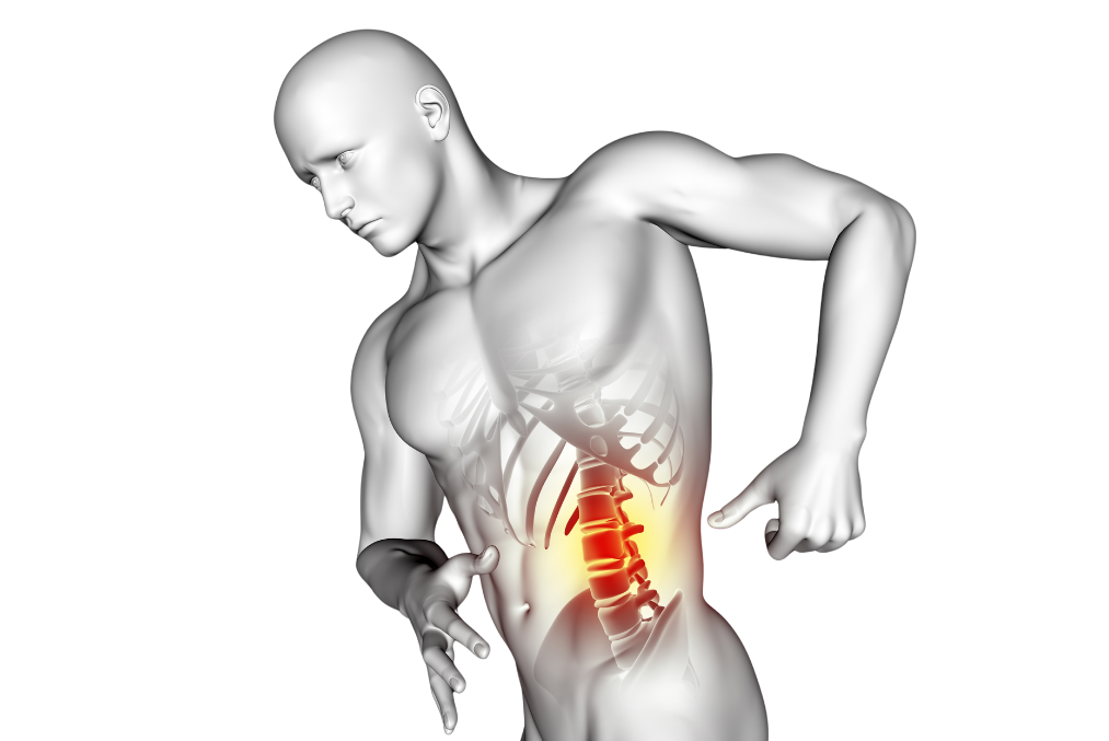
Spondylolisthesis, known among the public as "slipped disc," is more common in the lower part of the spine, particularly at the L4-L5 and L5-S1 vertebral levels. Slippages at these levels most frequently occur in the lumbar region, which carries the weight of the upper body. Especially heavy lifting, sudden movements, or traumas can disrupt the positions of the vertebrae and increase the pressure on the nerves.
Spondylolisthesis is not merely seen as a mechanical issue; this alteration in the spinal structure can also affect nerve functions, leading to serious neurological symptoms. Patients may experience severe pains, numbness, and muscle weakness, especially in the lower back and legs.
Anatomy and Physiology of Lumbar Spondylolisthesis
The spine is a structure consisting of 33 vertebrae extending from the lower part of the head to the coccyx. These vertebrae form the spinal canal, thereby protecting the spinal cord. The primary functions of the spine are to provide support to the body, offer mobility, and protect the nervous system. The lumbar region (lumbar area) is the part of the body that bears the most weight and contributes the most to mobility. Especially the L4, L5, and S1 vertebrae bear the body weight and support movements in the lumbar region.
How Does Spondylolisthesis Occur?
The spine is a structure that normally needs to maintain a certain balance. However, weakening of the discs between the vertebrae, wear of the spinal joints, or congenital structural disorders can cause the vertebrae to slip out of place. This slippage disrupts the overall stability of the spine and puts pressure on the nerve roots. The discs between the vertebrae, although crucial for enabling spinal mobility and protecting the vertebrae, can wear out or weaken over time. This leads to the slippage of the vertebrae.
The most critical factor in the process of spinal slippage is the loss of function in the spinal joints and intervertebral discs. Normally, these joints control the movement of the vertebrae and keep them together. However, degeneration or weakening of these structures can lead to the slippage of the vertebrae. This slippage can cause neurological symptoms by exerting pressure on the nerve roots. This pressure can lead to problems such as pain, numbness, and muscle weakness in the nerves.
When viewed in terms of physiological effects, the displacement of the vertebrae can increase pressure on the spinal cord and nerve roots, leading to problems in nerve communication. This pressure impedes the proper functioning of the nerves and leads to motor or sensory disorders. As the slippage of the lumbar region progresses, the load balance on the spine becomes uneven, and pain and discomfort can be felt in other areas of the body.
Causes of Lumbar Spondylolisthesis
There can be various causes for lumbar spondylolisthesis. These causes include congenital structural abnormalities, degenerative changes in the spine, traumas or injuries. In some cases, genetic factors can also increase the risk of lumbar spondylolisthesis. Below, the primary causes of lumbar spondylolisthesis are detailed.
Traumas and Injuries
Traumas are a common cause of spondylolisthesis. Direct impact on the spine, accidents, or sports injuries can lead to the displacement of the vertebrae. Particularly in athletes engaged in heavy sports, the slipping of vertebrae is more common due to sudden movements or muscle strains. The spine can lose its stability from its weak points against such sudden movements, leading to the displacement of the vertebrae.
High-impact sports (such as wrestling, football, weightlifting) and traumas like falls can cause damage to the spinal joints or discs. These types of injuries can disrupt the stability of the vertebrae and increase the symptoms of spondylolisthesis by exerting pressure on the nerve roots.
Degenerative Processes
Aging-related natural wear and tear on the spine is another factor that increases the risk of spondylolisthesis. The loss of water content and elasticity in the discs between the vertebrae leads to the weakening of the spinal joints over time. This, in turn, causes the vertebrae to slip out of place.
Degenerative spondylolisthesis arises due to wear and tear in the spinal joints and discs as a result of the aging process. With aging, the wear in the facet joints increases the mobility of the vertebrae, leading to their displacement. This process disrupts the natural structure of the spine and causes lumbar spondylolisthesis.
Genetic Factors
Genetic predisposition is a significant factor that increases the risk of spondylolisthesis. Individuals with a family history of spondylolisthesis may have a genetically weak spinal structure, which can make them more prone to slippage of the vertebrae. Especially, congenital abnormalities in the spinal structure can make it difficult for the vertebrae to align properly.
Congenital anomalies can cause the vertebrae to not develop properly due to disorders occurring during the development of the spine in some individuals. This situation is one of the primary causes of congenital spondylolisthesis cases. Particularly, developmental defects of the vertebrae in the lumbar region can disrupt the stability of the vertebrae, leading to slippage.
High Physical Stress
Occupations that require continuous heavy lifting or excessive physical activities can impose excessive strain on the spine, leading to spondylolisthesis. Especially in cases where the muscles are insufficient, the excessive loading on the spine causes the vertebrae to slip. High physical stress exceeds the load-bearing capacity of the spine, leading to the deterioration of joint and disc structures. Conditions such as heavy lifting and standing for long periods can cause the vertebrae to slip over time.
Types of Spinal Slippage
Lumbar spondylolisthesis, which refers to the displacement of a vertebra in the spine, can develop due to various reasons related to structural abnormalities in the spine. The classification of vertebral slippage depends on its cause and the degree of slippage, with each type based on different pathological processes. The treatment approaches vary according to these types. Determining the specific type of lumbar spondylolisthesis is critically important for the accurate formulation of a patient's treatment plan. Below, the most common types of lumbar spondylolisthesis are detailed.
Type 1: Dysplastic (Congenital) Spondylolisthesis
Dysplastic spondylolisthesis is a type of lumbar slippage that arises due to congenital structural abnormalities of the spine. Developmental anomalies of the vertebrae prevent the vertebrae from aligning properly, which in turn compromises spinal stability. The incomplete formation of the facet joints or the congenital weakness of the spinal bones plays a significant role in the development of dysplastic spondylolisthesis.
- Development Process: Dysplastic spondylolisthesis typically begins to develop during childhood or adolescence. As the spine develops, misalignment of the vertebrae or unstable joint structures can lead to vertebral slippage over time. The most common region for this condition is the end of the lumbar vertebrae, at the L5-S1 vertebral level. Weakening of the structures between the vertebrae in this area triggers the slippage.
- Symptoms: Dysplastic spondylolisthesis may not be detected at an early age. During childhood, the slippage of the vertebrae may be mild and symptoms may remain mild due to the absence of significant nerve compression. However, as age progresses and physical activity increases, the slippage becomes more pronounced. Especially during puberty, with the growth of bones, the slippage increases and symptoms become more severe. In patients with this type of disease, symptoms such as lower back pain, limited mobility, and muscle weakness typically emerge. With the increase in nerve compression, pain spreading to the legs may also be observed.
Type 2: Isthmic (Stress Fracture-Related) Spondylolisthesis
Isthmic spondylolisthesis is a type of lumbar slippage that develops as a result of weakening or fracturing of the bone structure called the pars interarticularis in the posterior part of the spine. These fractures typically occur due to repeated traumas, stress, or strains on the spine. The fractures in the pars interarticularis region lead to the forward slippage of the vertebrae over time. It is commonly seen in individuals who engage in heavy sports and participate in intense physical activities.
- Development Process: Isthmic spondylolisthesis typically emerges at a young age and develops as a result of excessive physical activities. It is more common among individuals engaged in activities that exert a lot of stress on the spine, especially athletes, football players, wrestlers, and weightlifters. After stress fractures occur in the vertebral bones, over time, the posterior part of the vertebrae weakens, leading to an anterior [forward] displacement. Non-healing of the fracture or repeated strains causing the fracture to enlarge can further advance the displacement.
- Symptoms: In the early stages, pain may be mild and is generally noticed during sports or strenuous physical activities. As stress fractures progress over time, spondylolisthesis becomes more pronounced and exerts pressure on the nerve roots, leading to severe pain. Patients often complain of symptoms such as lower back pain, pain radiating to the legs, numbness in the legs, and muscle weakness. In the advanced stages of the disease, neurological symptoms such as difficulty walking and loss of balance can also develop.
Type 3: Degenerative (Age-Related) Spondylolisthesis
Degenerative spondylolisthesis arises as a result of the wear and weakening of the disc and joint structures in the spine along with the aging process. Over time, the wear of the intervertebral discs and facet joints disrupts the stability of the vertebrae, leading to their slippage. Degenerative spondylolisthesis is typically observed in older individuals and develops as a consequence of the natural aging process of the spine.
- Development process: With aging, the discs between the vertebrae lose their water content and elasticity. This weakens their supportive role between the vertebrae. The degeneration of the facet joints also increases the mobility of the vertebrae, leading to slippage. Particularly in individuals over the age of 50, displacement of the vertebrae relative to each other is more common. Degenerative processes are generally more frequently observed at the L4-L5 vertebral level of the spine.
- Symptoms: Degenerative spondylolisthesis typically presents with lower back pain. Thinning of the intervertebral discs can lead to pressure on the nerve roots, causing sciatica-like pain. Patients may experience symptoms such as pain spreading from the lower back to the legs, numbness and weakness in the legs. In advanced cases, balance problems and difficulty walking are also common symptoms. Patients may struggle with standing and walking for extended periods.
Type 4: Traumatic Spondylolisthesis
Traumatic spondylolisthesis develops as a result of the spine being directly exposed to trauma, impacts, or accidents. Severe accidents, falls, or sports injuries can lead to fractures in the vertebral bones and damage to the ligamentous tissues. These traumas can cause the vertebrae to displace and their stability to be compromised.
- Development Process: Traumatic spondylolisthesis typically develops following a sudden trauma. Excessive load on the spine can cause fractures in the spinal bones. Fractures of the spine and damage to the ligamentous tissues can lead to the vertebrae slipping forwards or backwards. This type of displacement usually occurs after severe traumas and requires prompt intervention.
- Symptoms: Traumatic spondylolisthesis typically presents immediately after an accident with severe lower back pain. The slippage of the vertebrae can exert pressure on the nerve roots, leading to pain spreading to the legs, tingling, muscle weakness, and restricted movement. Patients may also notice spinal deformations and changes in posture. This type of slippage usually requires emergency intervention.
Type 5: Pathological Spondylolisthesis
Pathological spondylolisthesis develops as a result of tumors, infections, or other diseases affecting the bone tissue or connective tissues in the spine. These diseases can weaken the bone structure, leading to a loss of stability in the vertebrae and causing them to slip. Pathological processes impair the durability and structural integrity of the vertebrae.
- Development Process: Pathological spondylolisthesis develops due to bone diseases such as infections in the spine, bone tumors, or osteoporosis. These conditions weaken the bone structure and lead to the slipping of the vertebrae. Pathological processes usually emerge during the treatment of other underlying diseases. Osteoporosis is a disease that reduces the resilience of the spine and decreases bone density, which can lead to pathological spondylolisthesis.
- Symptoms: Pathological spondylolisthesis is often accompanied by general symptoms caused by abnormalities in the bone structure. Spinal pain, numbness in the legs, loss of strength, and neurological signs are common symptoms of the disease. Additionally, systemic symptoms such as fever, weight loss, and general malaise may also accompany, depending on the effects of a tumor or infection.
Grading of Slippage (Spondylolisthesis Grading)
The grading of spondylolisthesis is an important criterion that indicates how much the vertebra has slipped and plays a significant role in treatment planning. To measure how much the vertebra has displaced, the positions of the vertebrae are evaluated, and classification is made according to the amount of slippage. Cases of spondylolisthesis are divided into five different degrees based on the rate of slippage of the vertebra.
- Grade 1: A slippage of one vertebra over another by 25%. In these cases, symptoms are generally mild and conservative treatment methods (physical therapy, medications) may be sufficient.
- Grade 2: Slippage of the vertebra between 25% and 50%. Moderate pain, numbness in the legs, and balance issues may be observed. Physical therapy and medication are recommended, however, surgical options may become relevant in advanced cases.
- Grade 3: Slippage of the vertebra between 50% and 75%. It presents with severe pain, weakness in the legs, and serious neurological symptoms. Surgical treatment is generally recommended in these cases.
- Grade 4: Slippage of the vertebra between 75% and 100%. These cases are associated with severe spinal deformities. In these situations, where spinal stability is completely compromised, surgical intervention becomes inevitable.
- Grade 5 (Spondyloptosis): Cases where the vertebra is completely dislocated and the spinal canal is severely narrowed. This type of slippage is rare, but requires urgent surgical intervention.
Symptoms of Lumbar Spondylolisthesis
Lumbar spondylolisthesis, a condition characterized by the displacement of vertebrae, manifests through various symptoms due to the pressure exerted on nerve roots. Symptoms vary depending on the severity of the slippage, the direction in which the vertebra has slipped, and the extent to which the nerves are affected. While some patients may experience mild symptoms, others may suffer from severe pain and restricted movement. Below, the most common symptoms of lumbar spondylolisthesis are explained in detail.
Pain
Pain is the most common symptom of lumbar spondylolisthesis and usually begins in the lumbar region. The normal relationship between the vertebrae is disrupted as a result of the slippage, and this condition exerts pressure on the nerve roots, leading to pain. The pain in the lumbar region, which occurs together with the anterior or posterior displacement of the vertebrae, often radiates to the legs.
- Low Back Pain: The low back pain caused by spondylolisthesis intensifies over time due to increased pressure on the nerve roots. This pain becomes more pronounced especially when sitting, standing, or bending forward. Patients may feel more intense pain when getting out of bed in the morning or trying to move after sitting for a long period. The slippage of the vertebrae causes the muscles to tense, which in turn increases the pain.
- Pain spreading to the legs: When pressure is applied to the nerve roots exiting the spine, the pain is not limited to just the lower back area, but can spread down to the legs and feet. Particularly when the sciatic nerve is under pressure, patients may experience severe pain that starts from the lower back and extends down to the hips and legs. Sciatic pain, according to patients' descriptions, can feel like "burning", "electric shock", or "stabbing". This pain can especially intensify during activities such as walking, descending stairs, or sitting.
Numbness and Tingling
Lumbar spondylolisthesis can also lead to symptoms of numbness and tingling due to the pressure on the nerve roots. These symptoms arise from the compression of nerves that run along the spine. When nerves are under pressure, nerve transmissions are disrupted, resulting in numbness, tingling, or a sensation of pins and needles in the legs, hips, or feet.
- Numbness in the legs and feet: In patients with spondylolisthesis [slippage of a vertebra], numbness in the legs or feet can occur due to nerve compression. This condition is usually unilateral and varies depending on the direction of the vertebral slippage. Particularly during prolonged sitting or standing, this sensation can become more pronounced. Patients may describe a "pins and needles" sensation in their legs and may say that they cannot fully feel their legs.
- Tingling in the feet: Compression of the nerves can lead to a sensation of tingling that extends down to the feet. This condition intensifies with increased pressure on the nerve roots. Tingling results from insufficient neural stimulation reaching the leg muscles and can persist for the duration of the nerve compression.
Muscle Weakness
Muscle weakness is a common symptom in the advanced stages of spondylolisthesis. The spinal nerve roots control the movements of the leg muscles. When continuous pressure is applied to the nerve roots, the nerve signals to the muscles weaken, leading to muscle weakness. Muscle weakness can significantly limit the patient's mobility.
- Weakness in leg muscles: As nerve compression increases, loss of strength occurs in the leg muscles. Patients feel weakness in their legs while walking or climbing stairs. In some cases, patients report that their legs suddenly become weak and they experience a risk of falling. Muscle weakness originates from nerves being under pressure for an extended period and can become permanent if not treated.
- Foot Drop: When the nerves that control the upward movement of the ankle are compressed, a condition known as foot drop can develop. This condition makes it difficult for the patient to lift their foot and causes them to drag their foot while walking. Foot drop occurs in cases where nerve compression is severe and may require immediate intervention.
Movement Restriction
Spinal slippage disrupts the balance and mobility of the spine. The displacement of the vertebrae reduces the flexibility of the spine and limits mobility. The restriction of movement becomes more pronounced in cases where the spinal slippage progresses, causing patients to struggle with performing daily life activities.
- Difficulty in bending and twisting: Spondylolisthesis restricts the normal range of motion of the spine. Patients experience pain while bending, twisting, or turning their lower back, and their mobility decreases. This limitation in the lower back becomes a significant issue, especially for individuals engaged in heavy labor or those with high physical activity levels.
- Difficulty in Walking and Sitting: Increased pressure on the spine also affects patients' gait. Individuals with spondylolisthesis may become unable to walk long distances. Especially as the pressure on the spinal nerves intensifies, a sensation of weakness in the legs may occur during walking. Additionally, severe pain in the lower back and legs can be felt while sitting, which may shorten the duration of sitting.
Spinal Deformity
Spinal deformity is a symptom seen in the advanced stages of spondylolisthesis. Displacement of the vertebra can cause deformities in the spinal structure. In advanced cases, patients may notice visible curvatures or hunchback in their spines.
- Kyphosis or spinal curvature: Advanced level spondylolisthesis [slippage of a vertebra] can disrupt the natural structure of the spine and lead to spinal curvatures. This condition results in a visible distortion in the patients' posture. Particularly, patients with advanced degree of slippage (Grade 3-4) in the spine may struggle to stand upright, and permanent deformities can occur in the spine.
Spinal deformity, if left untreated, can significantly impact the patient's daily life and also cause aesthetic discomfort. In this case, surgical intervention is usually required.
Diagnosis of Lumbar Spondylolisthesis
The accurate and definitive diagnosis of lumbar spondylolisthesis requires careful evaluation of the patient's complaints and the use of necessary imaging methods. The diagnostic process is critically important in determining the degree of spinal slippage, measuring the severity of pressure on the nerve roots, and forming the treatment plan for the disease. During diagnosis, physical examination, imaging tests, and when necessary, nerve conduction studies are used in conjunction.
Physical Examination and Patient History
The first step in diagnosing lumbar spondylolisthesis is taking the patient's history and conducting a comprehensive physical examination. The doctor inquires about the symptoms experienced by the patient, the intensity of the pain, and how it affects their daily life. Additionally, information about the patient's current activities and any past traumas or injuries is collected.
- Patient's History: The doctor learns when the pain started, under what circumstances it intensified, and during which activities it subsided. It is investigated whether the patient has experienced any situation in their work or sports life, such as constant heavy lifting, straining, or falling, that could lead to spondylolisthesis [a condition where one of the vertebrae slips forward over the one below it]. Additionally, the presence of a history of spinal diseases in the patient's family is also assessed.
- Physical Examination: During the physical examination, the doctor evaluates the structure of the spine and tests spinal mobility. The patient is asked to perform movements such as bending, twisting, or turning. Pain and limitations in movement experienced by the patient during these activities are observed. In addition, nerve functions are also assessed. Muscle strength, reflexes, and sensory functions are tested to determine if there is any compression on the nerve roots. The presence of weakness, numbness, or tingling in the patient's legs is checked.
Radiological Imaging Methods
In the diagnosis of spondylolisthesis, radiological imaging techniques are used to clearly see the slippage in the spinal structure and the pressure on the nerve roots. These imaging techniques help determine the position of the vertebrae relative to each other and measure the degree of slippage.
- X-ray: This is the most commonly used imaging method to observe shifts in the bone structure of the spine. X-ray is an effective method to determine the degree of spondylolisthesis [slippage of a vertebra] and to see how much the vertebrae have displaced. Using X-ray films, the doctor can examine the placement of the vertebrae, the direction of the slippage, and other structural abnormalities in the spinal bones. X-ray images taken from the side clearly show whether the vertebrae have slipped forward or backward.
- Flexion and extension radiographs: The patient's spinal mobility and stability are assessed with X-ray films taken while the patient flexes and bends their spine. These radiographs are important for determining the amount of vertebral slippage and how the slippage changes during movement.
- Magnetic Resonance Imaging (MRI): MRI is utilized to visualize the soft tissues and nerve roots in the spine in detail. The condition of the discs between the vertebrae, the severity of pressure on the nerve roots, and changes in the soft tissues around the spine can be clearly seen with MRI. MRI is the best method for determining whether there is pressure on the nerve roots due to slippage of the vertebrae. Additionally, narrowing in the spinal canal (spinal stenosis) and disc degeneration between the vertebrae can also be visualized with MRI.
- Advantages of MRI: MRI is one of the most detailed methods for demonstrating the effects on the spine and nerve roots. It is preferred for evaluating the extent of compression on nerve roots and nerve damage because it does not contain radiation and clearly shows soft tissue structures.
- Computed Tomography (CT): CT is used to examine the details of the spinal bone structure. It provides clearer and more detailed images compared to X-rays. Structural changes such as the shape of the vertebrae, fractures, or bone protrusions are detected with CT. Since CT creates three-dimensional images of the spinal bones, it more clearly demonstrates the mechanical structure of vertebral slippage.
- Advantages of CT: CT is an effective method for detailed examination of the bone structures in the spine. Fractures, cracks, or damages in the vertebrae can be clearly visualized with this method. By revealing the three-dimensional structure of the spine, more information can be obtained about the direction and degree of slippage.
Electromyography (EMG) and Nerve Conduction Studies
In cases of vertebral slippage, the pressure on the nerve roots can disrupt nerve functions. The ability of nerves to transmit signals to muscles may decrease. Electromyography (EMG) and nerve conduction studies are used to assess the functions of nerves.
- Electromyography (EMG): EMG measures the electrical activity of muscles. It is used to assess how effectively nerves are sending signals to the muscles. If the pressure on the nerve roots increases due to spinal slippage, the signals reaching the muscles weaken, and muscle weakness occurs. The EMG test can understand how the muscles are functioning and how the nerves affect these muscles. The degree of impairments in nerve functions is determined by this method.
- Nerve conduction studies: Nerve conduction studies measure how quickly nerves transmit electrical signals. These tests are used to detect disorders in nerve transmission. If there is pressure on the nerve roots, the response times of the nerves to stimuli in these conduction tests lengthen, and the conduction velocity decreases. Nerve conduction studies are important in determining the severity of nerve compression and the degree of loss in nerve functions.
Treatment Methods for Lumbar Spondylolisthesis
The methods used in the treatment of lumbar spondylolisthesis vary depending on the severity of the symptoms experienced by the patient, the degree of the slip, and the pressure on the nerves. The aim of the treatment is to alleviate pain, eliminate the pressure on the nerve roots, and restore the stability of the spine. Treatment options range from non-surgical methods (conservative treatment) to surgical interventions. Below, the methods used in the treatment of lumbar spondylolisthesis are explained in detail.
Conservative (Non-Surgical) Treatment Methods
In the early stages of the disease, if the degree of vertebral slippage is low (Grade 1-2), conservative treatment methods are applied as the first option. These methods are used to alleviate the pressure on the nerve roots, strengthen the muscles, and control the patient's pain. Symptoms of patients are significantly alleviated through lifestyle changes, physical therapy, and medication therapy.
```json
{
"li": {
"strong": "Physical Therapy and Rehabilitation:",
"text": "Physical therapy is one of the most common conservative treatment methods applied to strengthen the muscles supporting the spine of patients with spondylolisthesis and to increase their mobility. Strengthening especially the lumbar and abdominal muscles increases the stability of the spine and prevents the progression of the slip.",
"ul": {
"li": [
{
"strong": "Exercise programs:",
"text": "Exercise programs prepared by physical therapy specialists are designed to strengthen the muscles supporting the spine. These exercises provide additional support to the patient's spine and improve the mobility of the vertebrae. Exercises such as planks, bridge exercises, and other exercises that strengthen the back muscles are beneficial for patients with spondylolisthesis. Moreover, strengthening the lumbar and abdominal muscles reduces the pressure on the nerve roots."
},
{
"strong": "Flexibility and posture correction:",
"text": "Physical therapy also includes stretching movements aimed at correcting patients' posture and increasing the flexibility of the spine. Incorrect posture can apply additional pressure on the spine, exacerbating the slip, hence proper posture habits are encouraged."
}
]
}
}
}
``` ```json
{
"Drug Therapy": "- Drug Therapy: Various drug therapies can be applied to control the pain and inflammation caused by spondylolisthesis. Medications alleviate pain and reduce muscle spasms, enabling the patient to maintain a more comfortable daily life.
- Non-steroidal anti-inflammatory drugs (NSAIDs): These medications are used to relieve pain and inflammation resulting from spondylolisthesis. NSAIDs such as ibuprofen and naproxen reduce inflammation caused by pressure on nerve roots and have analgesic [pain-relieving] effects. These drugs are especially preferred for mild to moderate pain.
- Muscle relaxants: Spondylolisthesis can cause muscle spasms, which in turn increase pain. Muscle relaxants help relax the muscles, thereby easing spasms. Medications like tizanidine or baclofen can reduce muscle tension in the lumbar region, enhancing the patient's mobility.
- Pain relievers: For patients experiencing severe pain, stronger pain relievers such as opioids (morphine, tramadol) may be recommended. However, these medications carry a risk of addiction and are generally used for a short duration. Pain relief treatment is reduced as the pressure on the spine decreases and other treatment methods are initiated.
"
}
``` - Braces and Support Devices: In some cases, braces or supportive devices are used to control lumbar spondylolisthesis and ensure the spine is more stable. These devices help to immobilize the spine and reduce the load on the lumbar region during the patient's daily activities.
- Use of braces: Braces assist in maintaining the correct posture of the spine and prevent the progression of vertebral slippage during movement. Especially when a sudden load is applied to the spine, braces provide support to the lumbar region and alleviate the patient's pain. However, since prolonged use of braces can lead to muscle weakening, they should only be used with a doctor's recommendation.
- Corticosteroid Injections: In patients experiencing severe pain due to compression of nerve roots, corticosteroid injections can be utilized. These injections reduce inflammation around the spine, thereby temporarily alleviating pain. Corticosteroids are injected directly into the tissues surrounding the nerve roots, easing inflammation and reducing nerve compression.
- Epidural Steroid Injection: Corticosteroids are injected into the epidural space surrounding the spinal cord. This allows the nerve roots to relax and results in a significant reduction in the patient's pain. However, this treatment method generally provides temporary relief and its use in conjunction with other treatment methods is recommended.
Surgical Treatment Methods
Lumbar spondylolisthesis, a condition where the vertebrae slip out of place, results in the loss of spinal stability and can lead to serious neurological symptoms by exerting pressure on the nerve roots. If conservative (non-surgical) treatment methods prove insufficient and the patient's complaints progress, surgical treatment options may be considered. The goal of surgical intervention is to stabilize the slipped vertebrae, relieve the pressure on the nerve roots, and restore spinal stability. Below, the most commonly applied surgical methods for the treatment of lumbar spondylolisthesis are discussed in detail.
Spinal Fusion
Spinal fusion is a surgical procedure performed to provide stability to the spine and fix slipped vertebrae. This process is accomplished by fusing the slipped vertebrae together, thereby eliminating abnormal movement between the vertebrae. Spinal fusion is generally preferred in cases of advanced vertebral slippage and severe nerve compression.
```json
{
"How is fusion performed?": "During spinal fusion, the surgeon uses various techniques to fix the vertebrae together. To increase the stability of the vertebrae, metal plates, screws, or rods are inserted. These stabilizing devices stop the vertebrae from slipping, ensuring they stay in the correct position. Bone grafts are also used during the fusion process; these grafts can be taken from the patient's own bone tissue, synthetic materials, or bone tissues obtained from cadavers. Over time, these bone grafts create new bone tissue between the vertebrae, preventing slippage and completely fusing the vertebrae together. The spinal fusion procedure eliminates abnormal movement in the slipped part of the spine, thereby relieving pressure on the nerve roots."
}
``` - Advantages of Fusion: Spinal fusion is an extremely effective method in providing spinal stability. Post-surgery, patients generally experience a significant decrease in lower back pain. Since the pressure on the nerve roots is eliminated, neurological symptoms (numbness, tingling, and weakness in the legs) also improve greatly. Moreover, once the slipped vertebrae are stabilized, deformities in the spine are also corrected. Patients can walk more comfortably after the surgery, and their mobility is significantly restored.
- Disadvantages of Fusion: Among the disadvantages of spinal fusion is the restriction of mobility in the spine. Since the vertebrae are fused together, movement in this area is completely eliminated, which can create mobility restrictions for some patients. The recovery time after the fusion procedure can be quite long; the complete healing process can take months, and patients may need physical therapy. Additionally, due to the increased load on other areas of the spine, degenerative problems can develop in other regions of the spine over time.
Decompression Surgery
Decompression surgery is a surgical intervention applied to eliminate the pressure exerted on the nerve roots. Narrowing in the spine (spinal stenosis) due to slippage of the vertebrae can cause pain spreading to the legs, numbness, and muscle weakness by putting pressure on the nerve roots. Decompression surgery encompasses various surgical techniques that expand the spinal canal to alleviate this nerve pressure.
Laminectomy
Laminectomy is a decompression surgery performed by removing a bone structure called the lamina, which is located at the back part of the spine. The lamina is situated at the back of the spinal canal and protects the spinal cord and nerve roots. However, in the case of spondylolisthesis [slippage of one vertebra over another], this structure can press on the nerve roots, therefore, through the laminectomy procedure, this bone structure is removed and the spinal canal is widened.
- How is a laminectomy performed?: The surgeon makes an incision over the spine and removes a bone called the lamina. This procedure relieves the area where the nerve roots are compressed and expands the spinal canal, allowing the nerves to be freed. Laminectomy is usually preferred in cases of spinal canal narrowing, such as spinal stenosis [a condition characterized by the narrowing of the spaces within your spine, which can put pressure on the nerves that travel through the spine], and is used in cases of severe nerve compression. Since the pressure on the spinal cord is eliminated with laminectomy, patients generally feel rapid relief after the surgery.
- Advantages of Laminectomy: Laminectomy effectively relieves pressure on the nerve roots by widening the spinal canal. Patients feel a significant relief in their legs and lower back regions immediately after the surgery. Moreover, this method significantly restores walking capacity, especially in patients who experience difficulty walking due to nerve compression.
Laminotomy
Laminotomy is a procedure similar to laminectomy, but it is applied as a more minimally invasive technique. During laminotomy, the lamina is not completely removed; instead, only a part of it is taken to widen the spinal canal. This method creates less tissue damage and the recovery time may be shorter.
- How is a laminotomy performed?: During a laminotomy procedure, instead of completely removing the lamina, which is a bony structure, only the part exerting pressure is cut to expand the spinal canal. This process alleviates pressure on the spine while preserving more of the spine's integrity. Laminotomy relieves pressure on the nerve roots and aids in freeing the nerves without compromising the stability of the spine.
- Advantages of Laminotomy: Laminotomy is a less invasive method compared to laminectomy, hence the recovery time is shorter. Since it causes less damage to the tissues on the spine, patients recover faster after surgery. Moreover, since the stability of the spine is largely preserved, the restriction on spinal mobility is minimized. Therefore, laminotomy is preferred in milder cases or in patients where nerve compression is relatively less severe.
Minimally Invasive Surgical Techniques
Minimally invasive surgical techniques are procedures performed with small incisions in spine surgery. These methods create less tissue damage compared to traditional surgeries, accelerate the healing process, and minimize the risk of complications. Minimally invasive surgeries can be effectively applied in cases of spondylolisthesis [slippage of one vertebra over another] and nerve compression. They allow patients to return to their normal activities in a shorter period of time.
Microdiscectomy
Microdiscectomy is a minimally invasive technique, performed with the aim of removing a small portion of the herniated disc that is pressing on the nerve roots. This procedure is preferred in cases of spondylolisthesis, where the discs shift between the vertebrae and press on the nerves.
- How is a microdiscectomy performed?: The surgeon makes a small incision on the spine with the aid of a microscope to remove the portion of the herniated disc that is pressing on the nerve roots. During this procedure, other healthy tissues are not damaged, and the pressure on the nerve roots is quickly eliminated. Since microdiscectomy is a minimally invasive method, the duration of the surgery is shorter, and patients can return to their normal activities shortly after the operation.
- Advantages of Microdiscectomy: Since microdiscectomy is a minimally invasive technique, the recovery time after surgery is quite short. As healthy tissues in the spine are not damaged during the surgery, patients can usually stand up quickly after the operation. Moreover, since it is performed with small incisions, the risk of infection, blood loss, and other surgical complications are reduced. Patients can return to their normal activities within a few weeks after the surgery.
Advantages of Minimally Invasive Surgical Techniques
Minimally invasive surgical techniques have numerous advantages compared to traditional open surgical methods:
- Smaller incisions: Minimally invasive surgeries are performed with smaller incisions on the spine, which accelerates post-surgical wound healing and reduces the risk of infection.
- Shorter recovery time: Since less damage is inflicted on the tissues in the spine, the recovery time is much shorter compared to traditional surgeries. Patients can usually return to their normal lives within a few weeks after the surgery.
- Reduced blood loss: Small incisions and less invasive techniques keep blood loss at a minimal level, which accelerates postoperative recovery.
- Less pain: Following minimally invasive surgical techniques, patients experience less pain, which leads to a more comfortable recovery process after surgery.
Complications and Long-Term Outcomes of Lumbar Spondylolisthesis
Lumbar spondylolisthesis, if left untreated or if it progresses, can lead to various complications due to nerve compression and disruption of spinal stability. These complications can significantly affect the patient's quality of life and can cause permanent damage. The severity of the complications depends on the degree of slippage, the duration of pressure on the nerve roots, and the timing of the treatment. Here are the most common complications associated with lumbar spondylolisthesis and the long-term effects of this condition:
```json
{
"Permanent Nerve Damage": "One of the most serious complications of spinal slippage is permanent nerve damage. The continuous pressure applied to the nerve roots due to the displacement of the vertebrae can cause the nerves to lose their function over time. This condition negatively affects the nerves' ability to send signals to muscles and sensory areas.",
"Symptoms of Nerve Damage": [
{
"Symptoms": "As a result of prolonged nerve compression, patients may experience weakness, numbness, tingling, and loss of reflexes in the legs. In advanced cases, nerve damage can become permanent, and the patient's mobility can be severely restricted. The recovery of nerve functions can become more difficult depending on the duration of the pressure and the degree of damage in the nerves."
}
],
"Treatment of Nerve Damage": [
{
"Treatment": "Nerve damage is generally irreversible. If nerve compression has continued for a long time, permanent damages can occur in the nerve cells. In such cases, treatment may be aimed at managing muscle weakness and sensory losses, depending on the degree of nerve damage."
}
]
}
``` ```json
{
"li": {
"strong": "Disruption of Spinal Stability:",
"text": "The progression of spondylolisthesis can lead to the disruption of spinal stability. The slippage of vertebrae disrupts the balance of the spine, which can cause structural abnormalities in other parts of the spine. Disruption of spinal stability can negatively affect the patient's ability to walk and can lead to severe postural disorders.",
"ul": {
"li": [
{
"strong": "Symptoms of spinal stability loss:",
"text": "The loss of stability in the spine affects the patient's posture and causes the normal alignment of the spine to be disrupted during walking. This condition can lead to deformities such as kyphosis [an excessive outward curvature of the spine, resulting in a hunchback appearance] in patients. The loss of balance in the spine can lead to continuous muscle spasms and pain."
},
{
"strong": "Treatment options:",
"text": "Restoring spinal stability usually requires surgical intervention. Spinal fusion or other surgical procedures aimed at increasing the stability of the spine can help in fixing the vertebrae. After treatment, patients may need regular physical therapy to increase the stability of the spine."
}
]
}
}
}
``` - Muscle Weakness and Loss of Movement: Lumbar spondylolisthesis compresses the nerve roots, blocking the nerve signals to the muscles. This condition can lead to weakening of the leg muscles and prevent the patient from performing daily activities. Due to the disruptions in nerve functions, patients experience muscle weakness and loss of movement, especially in their legs.
- Muscle weakness: As nerve compression increases, it weakens the nerve signals reaching the muscles. This condition leads to weakness and movement disorders in the legs. A condition known as foot drop, where the patient cannot lift their foot, may result in dragging the foot while walking. As muscle weakness progresses, the patient's ability to move can be further restricted.
- Loss of movement: If muscle strength is lost due to nerve damage, patients may struggle to perform daily movements. Simple actions like climbing stairs, walking, or standing can become difficult. Loss of movement is generally dependent on the duration the nerves have been under pressure and can become permanent if not treated.
- Chronic Pain: When spondylolisthesis is not treated, chronic pain can develop due to the pressure on nerves and disturbances in the stability of the spine. The pain becomes constant due to structural abnormalities in the spine and the compression of nerve roots. Chronic pain can significantly affect the quality of life of patients and may require long-term treatment.
- Chronic Lower Back Pain: Patients with spondylolisthesis often experience severe and constant pain in the lower back region. This pain stems from the misalignment of the spine and intensifies with physical activities. Prolonged sitting, standing, or bending movements can trigger the pain.
- Pain Spreading to the Legs: The nerve compression resulting from spondylolisthesis can cause pain that spreads down to the legs. Sciatic nerve pain, starting from the lower back and extending down to the hips and legs, can lead to sharp and burning sensations. This pain makes it difficult for patients to move and restricts their daily activities.
- Loss of Bladder and Bowel Control: As spondylolisthesis progresses and severe pressure is applied to the spinal nerves, patients may develop serious neurological complications such as loss of bladder and bowel control. Particularly, a condition known as cauda equina syndrome requires immediate medical intervention.
- Cauda Equina Syndrome: This condition arises from severe compression of the nerve roots located at the lower part of the spinal cord. These nerves manage the motor and sensory functions of the legs, as well as the control of the bladder and bowels. Pressure on these nerve roots can lead to loss of bladder control, urinary incontinence, or loss of bowel control. Patients may experience sudden weakness in their legs, numbness, and tingling between the legs.
- Treatment Process: Cauda equina syndrome requires urgent surgical intervention. The goal of treatment for this syndrome is to quickly relieve the pressure on the nerve roots, thereby restoring nerve function. If the intervention is delayed, patients may suffer permanent loss of bladder and bowel control.
```json
{
"li": {
"strong": "Spinal Deformities:",
"text": "In advanced cases, spondylolisthesis can lead to structural disorders in the spine. This situation can cause permanent deformities in the spine and posture disorders. Especially in cases where spinal stability is severely compromised, patients can develop visible spinal curvatures.",
"ul": {
"li": [
{
"strong": "Kyphosis or Scoliosis:",
"text": "Among spinal deformities, the most common ones are known as kyphosis (hunchback) and scoliosis (lateral curvature). These deformities occur as a result of the disruption of spinal balance and the abnormal positioning of the vertebrae. Kyphosis causes the patient to bend forward, while scoliosis leads to lateral bending of the spine."
},
{
"strong": "Treatment Process:",
"text": "Spinal deformities generally require surgical intervention. Screws, rods, or other stabilizers are placed in the spine to correct the deformities and fix the spine in the correct position. After surgery, patients may need long-term physical therapy to maintain spinal stability."
}
]
}
}
}
```
Methods of Preventing Lumbar Spondylolisthesis
Lumbar spondylolisthesis, a condition characterized by the slippage of a vertebra, develops as a result of excessive load on the spine and a weak spinal structure. To prevent lumbar spondylolisthesis, it is crucial to maintain spinal health, strengthen muscles, and take certain precautions in lifestyle. This section will detail the preventive measures that can be implemented to prevent the development of lumbar spondylolisthesis and to maintain spinal health.
Proper Posture and Ergonomic Measures
Maintaining proper posture is one of the fundamental steps in preserving spinal health. A posture that supports the natural curvatures of the spine reduces the load on the lumbar and back region and decreases the risk of developing spondylolisthesis [a condition where one vertebra slips forward over the one below it]. Sitting or standing in an incorrect position for extended periods applies unnecessary pressure on the spine, and this pressure can lead to vertebral slippage over time.
-
Correct posture while sitting: The sitting position is of great importance, especially for people who work at desks. Using an ergonomic cushion that supports the natural curve of the spine while sitting in a chair reduces the load on the spine. Feet should be planted firmly on the ground, knees should be bent at a 90-degree angle, and the back should be kept straight. Having the computer screen at eye level prevents extra strain on the neck and back muscles. Additionally, one should avoid sitting for prolonged periods and take frequent breaks to stand.
-
Proper posture while standing: Standing for extended periods can exert pressure on the spine. Distributing weight evenly across both feet and keeping the shoulders back helps maintain the spine in the correct position. To prevent lumbar spondylolisthesis [slippage of a lumbar vertebra], it is beneficial to frequently change positions while standing and slightly elevate one foot (for example, placing it on a step), which preserves the balance of the spine.
-
Proper techniques during heavy lifting: One of the factors that increase the risk of spondylolisthesis [a condition where a vertebra slips forward over the bone below it] is incorrect movements while lifting heavy loads. Lifting weight by bending from the waist applies excessive pressure on the spine and can cause the vertebrae to slip out of place. When lifting weights, one should squat by bending the knees and keep the back straight. The weight should be kept close to the body and lifted with a slow movement. This technique reduces the risk of spondylolisthesis while protecting the spine.
Exercise and Physical Activity
Regular exercise plays a critical role in preventing lumbar spondylolisthesis. Having strong muscles that support the spine balances the pressure on the spine and prevents the vertebrae from slipping. Strengthening the lumbar, abdominal, and back muscles in particular increases spinal stability. Exercise programs not only strengthen the muscles but also increase the flexibility of the spine, providing protection against sudden strains.
-
Exercises that strengthen the abdominal and back muscles: Having strong muscles that support the spine is one of the most effective methods to prevent spondylolisthesis [a condition where a vertebra slips forward over the bone below it]. Exercises such as planks, bridge poses, and the superman exercise strengthen both the abdominal and back muscles. The strength of these muscles helps to keep the vertebrae stable and prevents slipping. Moreover, the balanced strengthening of muscles on both sides of the spine maintains its stability.
-
Stretching exercises: Keeping the spine flexible is another way to prevent lumbar spondylolisthesis. Yoga and Pilates are among the exercises that increase the flexibility of the spine and ensure muscle balance. These exercises, while increasing the mobility of the spine, also strengthen the muscles. A flexible spine is more resistant to sudden strains and loads.
-
Aerobic exercises: Low-impact aerobic exercises such as walking, swimming, and cycling are ideal for maintaining spinal health. Swimming, in particular, minimizes pressure on the spine while exercising the muscles and preserving joint health. Regular walking strengthens the muscles in the lumbar region while preventing excessive load on the spine.
Avoiding Heavy Lifting and Correct Lifting Techniques
One of the common causes of lumbar spondylolisthesis is improper weight lifting techniques. Lifting heavy loads incorrectly can increase the pressure on the spine, leading to vertebral slippage. Therefore, avoiding heavy lifting and applying correct lifting techniques is extremely important.
-
Avoid lifting heavy objects: Avoiding heavy loads as much as possible reduces the risk of spondylolisthesis. If it is necessary to lift a heavy load, this task should be performed by several people or the load should be divided and carried. Additionally, carrying loads for a long time or repeated heavy lifting should be avoided.
-
Proper lifting techniques: Lifting weights by bending the knees reduces the load on the spine. Instead of bending from the waist to lift weights, one should squat from the knees and keep the weight close to the body. This technique protects the muscles and joints in the lower back region and prevents the displacement of the vertebrae. Additionally, the lifted load should never be taken over the lower back; the load should be carried parallel to and close to the body.
Control of Body Weight
Obesity and overweight are significant factors that negatively affect spinal health. Excess weight places undue stress on the spine, and this load can damage the vertebrae over time. In particular, excess weight in the lumbar region can cause the vertebrae to slip over time.
-
Healthy weight range: Losing excess weight reduces the pressure on the spine and minimizes the risk of spondylolisthesis. Healthy eating and regular exercise are effective methods for weight control. A healthy body weight ensures the spine carries loads in a balanced manner and prevents the vertebrae from slipping.
-
Reducing fat accumulation around the waist: Fat deposits, especially in the waist area, impose an additional load on the spine and increase the risk of spondylolisthesis [a condition where a vertebra slips forward over the bone below it]. Strong abdominal muscles and a low fat ratio in the waist area help maintain the spine in its correct position. Low-carbohydrate diets and regular cardiovascular exercises support spinal health by reducing fat accumulation around the waist.
Cigarette Smoking and Spinal Health
Smoking has adverse effects on spinal health. Smoking prevents the discs between the vertebrae from receiving adequate oxygen and nutrients. The lack of nourishment for the discs leads to their weakening over time and loss of elasticity. This situation causes the discs to lose their function, thereby increasing the risk of spondylolisthesis [a condition where a vertebra slips forward over the bone below it].
-
The effect of smoking on discs: In individuals who smoke, the discs are not adequately nourished, and the flexibility of the discs decreases. This leads to quicker wear and tear of the discs and an increased risk of herniation. The acceleration of disc degeneration reduces the flexibility and durability of the spine.
-
Cessation of smoking: Quitting smoking facilitates the healthy nourishment and oxygenation of the discs. When smoking is ceased, the healing capacity of the discs increases, and there can be a significant reduction in the risk of spondylolisthesis. Thus, the health of the spine is preserved, and the likelihood of developing spondylolisthesis decreases.
Regular Doctor Check-ups
To prevent lumbar spondylolisthesis, it is important to regularly monitor spinal health. Especially individuals experiencing lower back and back pain should not neglect regular medical check-ups for spinal health. Early-stage examinations can be effective in preventing lumbar spondylolisthesis.
-
Early diagnosis and intervention: The early diagnosis of spondylolisthesis is extremely important in preventing the progression of the disease. Regular doctor visits play a critical role in maintaining spinal health and detecting structural abnormalities in the spine at an early stage.
-
Monitoring of Symptoms: Individuals at risk for spinal health (those working in jobs requiring heavy lifting, athletes, or those with a genetic predisposition) should undergo regular doctor check-ups. Exercise programs and lifestyle changes recommended by a doctor can reduce the risk of spondylolisthesis.
Result
Lumbar spondylolisthesis, despite being a serious condition that threatens spinal health, can be successfully managed with the correct diagnosis and appropriate treatment methods. This disorder manifests itself through symptoms such as lower back and leg pain, muscle weakness, and restricted movement, due to the displacement of vertebrae which puts pressure on the nerve roots. However, the symptoms caused by lumbar spondylolisthesis can be largely controlled with early diagnosis and effective treatment.
Early diagnosis is the most critical step in stopping the progression of spinal slippage. Patients should consult a specialist without delay when they notice symptoms such as pain, numbness, and weakness in their back and legs. Through the use of diagnostic methods such as physical examination, X-ray, MRI, and nerve conduction studies, the degree of slippage and the pressure on the nerve roots can be assessed in detail. This allows for the determination of the stage of the disease and the formulation of the most appropriate treatment plan.
Conservative (non-surgical) methods in the treatment of lumbar spondylolisthesis, especially in the early stages of the disease, yield effective results. Physical therapy strengthens the muscles and increases the stability of the spine, while medication therapy controls pain and inflammation. In addition, spinal support corsets and corticosteroid injections can also be used to improve the quality of life for patients. These methods are quite successful in stopping the progression of vertebral slippage and alleviating symptoms.
Surgical treatment methods are applied in cases that do not respond to conservative treatments or carry a risk of nerve damage. Spinal fusion, laminectomy, and minimally invasive surgical techniques are utilized to re-establish spinal stability and relieve pressure on the nerve roots. These surgical interventions enable patients to regain mobility and significantly reduce pain. Each surgical procedure is personalized according to the patient's condition and the severity of the slippage.
Lumbar spondylolisthesis is a condition that can be managed with appropriate treatment approaches. After treatment, the majority of patients can return to their normal daily activities and regain their quality of life. However, changes made in lifestyle to prevent recurrence of lumbar spondylolisthesis after treatment are of great importance. Regular exercise helps strengthen the muscles that support the spine and maintains spinal stability. Additionally, avoiding heavy lifting, adopting correct posture habits, and maintaining weight control minimize the risk of lumbar spondylolisthesis.
Simple measures to protect spinal health and prevent spondylolisthesis can largely prevent the occurrence of this condition. A healthy spine is of critical importance for the continuation of life quality. Especially individuals working in heavy labor, those engaged in sports, and people with genetic factors prone to spinal health should make protecting their spine a part of their life. Regular doctor check-ups can identify problems in the spine at an early stage, allowing for solutions without the need for surgical intervention.
In conclusion, for patients with spondylolisthesis, the critical aspects are early diagnosis, accurate treatment, and lifestyle changes that will preserve spinal health. It is important for patients to adhere to doctor recommendations by engaging in regular exercise and maintaining strong muscles that support the spine, especially during the post-treatment period. It is essential to remember that spondylolisthesis is treatable; with the right treatment plan, patients can be relieved of their pain and continue their lives in a healthy manner.