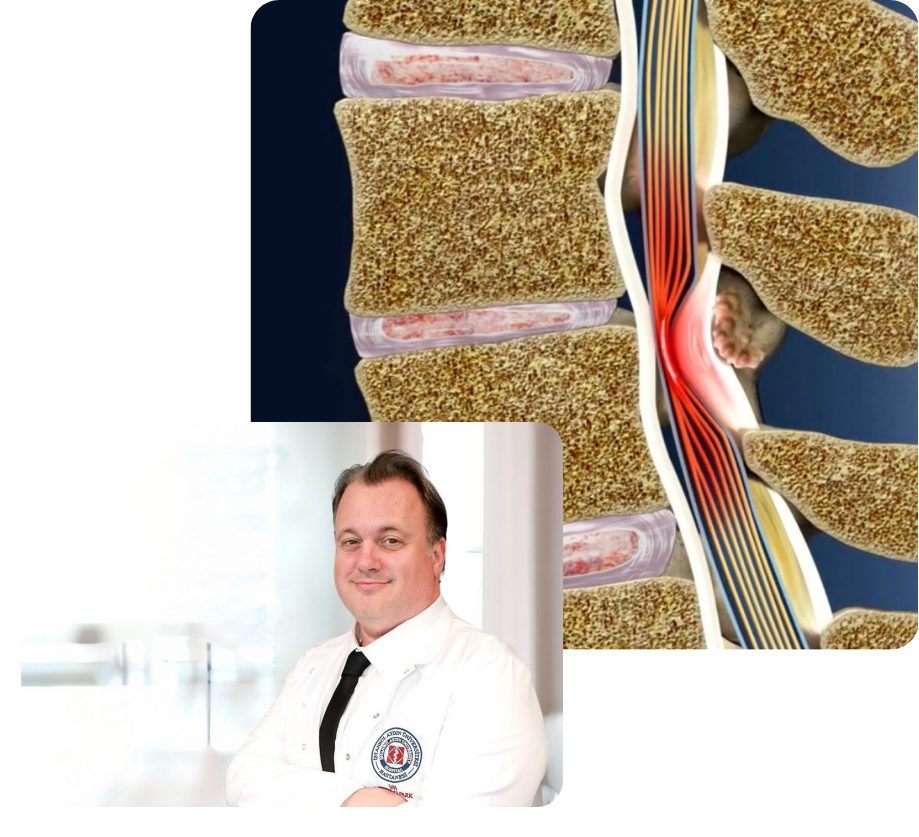
Narrowing of the spinal canal can cause damage to the spinal cord and nerve roots. This condition can lead to pain, numbness, and loss of movement in various parts of the body, primarily in the neck and arms. If not treated in a timely manner, cervical spinal stenosis can lead to neurological complications and significant declines in quality of life.
The effects of spinal stenosis in the neck generally develop gradually and can make it difficult for the patient to move and perform daily activities as it progresses. Disruptions in the function of the cervical vertebrae can lead to serious neurological complications due to the interruption of nerve transmission.
What Are the Symptoms of Cervical Spinal Stenosis?
Cervical spinal stenosis occurs due to the narrowing of the spinal canal, resulting in pressure on the nerve roots and spinal cord. Symptoms vary depending on the degree of narrowing and the intensity of pressure applied to the spinal cord and nerve roots. Initially, it may present with mild symptoms and can lead to more serious neurological symptoms as it progresses over time. Here are the most common symptoms of cervical spinal stenosis:
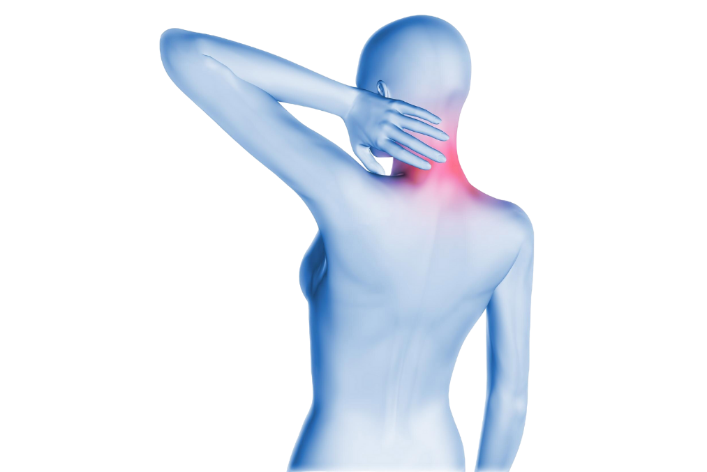
Neck Pain
Neck pain is one of the most common and early symptoms of cervical spinal stenosis. The pain originates from the nerves and spinal cord in the neck being under pressure and is usually described as a constant, uncomfortable sensation. The intensity of the pain can vary from patient to patient; while some may experience mild discomfort, for others, it can be severe and persistent. The pain may increase during neck movements and can make daily activities difficult.
- Prevalence: Neck pain is usually felt in a constant manner, but it can increase with sudden head movements or bending of the neck. This condition can negatively affect the patient's quality of life.
- Character of the pain: The pain can be described as a constant discomfort, while in some patients, a burning or sharp pain may also be observed. The pain can intensify with the patient's turning or stretching of the neck.
Numbness and Tingling in the Arms and Hands
One of the common symptoms of cervical spinal stenosis is numbness and tingling in the arms and hands. These symptoms occur due to the compression of nerve roots caused by the narrowing of the spinal canal. Numbness is usually felt in the arms, hands, and fingers and can worsen with increased pressure on the nerve roots. Particularly when direct pressure is applied to the nerve roots, patients may struggle to use their hands.
- Affected areas: Numbness and tingling felt in the arms, hands, and fingers are caused by disturbances in nerve conduction. These symptoms can particularly increase with staying in the same position for a long time.
- Perceived changes: Numbness and tingling are usually associated with a decrease or distortion in sensation. This condition arises as a result of the nerves not being able to transmit signals adequately.
Weakness in the Hand and Fingers
Weakness in the hands and fingers is a serious symptom that emerges with the progression of cervical spinal stenosis. Prolonged compression of the nerve roots can disrupt motor functions, leading to weakening of the muscles in the hands and fingers. Patients may experience weakness in their hands and difficulty performing daily tasks. Actions requiring fine motor skills, such as writing, buttoning, or grasping an object, may become more difficult.
- Strength loss: Weakness in the hand and finger muscles can lead to difficulties in performing simple tasks such as gripping or holding objects. This condition can significantly affect the patient's daily activities.
- Impairment in fine motor skills: Difficulties may be observed in tasks requiring hand dexterity (buttoning, writing). These symptoms can progress over time, further deteriorating the patient's hand skills.
Additional Symptoms Resulting from Compression of Nerve Roots
Pressure on the nerve roots becomes more pronounced with the progression of cervical spinal stenosis, and this pressure can cause symptoms in other areas of the body as well as in the arms. As a result of the nerves emanating from the spinal cord being under pressure, patients may experience widespread pain, muscle weakness, and loss of balance in the areas where the nerve roots spread.
- Pain in the arms and shoulders: As a result of the nerve roots being affected, pain can spread not only to the neck but also to the arms and shoulders. Compression of the nerves can cause a burning or stabbing type of pain.
- Muscle weakness: Continuous pressure applied to the nerve roots leads to the weakening of muscles and loss of strength in these areas. As muscles weaken, the patient may struggle to use their arms and legs.
- Balance loss: Damage to the nerve roots can also affect the body's balance. Patients may experience a sense of imbalance, especially while walking.
Neurological Symptoms
As cervical spinal stenosis progresses, the pressure on the spinal cord increases, leading to neurological symptoms. Prolonged pressure on the nerve roots and spinal cord can result in severe symptoms that spread to the lower regions of the body. Neurological symptoms can lead to permanent damage if not treated.
- Weakness in the legs: In advanced stenosis cases, the pressure on the spinal cord can extend to the legs, causing muscle weakness. Patients may experience difficulties in walking, inability to lift their feet off the ground, and similar issues.
- Walking difficulties: Pressure on the spinal cord can affect nerve conduction in the legs. This condition can make it difficult for the patient to walk, cause imbalance while taking steps, and make it challenging to perform daily tasks.
- Decrease in reflexes: With the intensification of neurological symptoms, patients' reflexes may also weaken. Reflex loss refers to a reduction in the body's responses to external stimuli.
- Coordination disorder and loss of balance: In advanced cases, compression of the spinal cord can lead to coordination problems, and the patient may experience loss of balance during daily activities. This situation can cause falls and injuries.
Additional Symptoms Observed in Advanced Stenosis
In advanced cases of cervical spinal stenosis, the pressure on the spinal cord and nerve roots can lead to serious complications. Damage to the spinal cord, known as myelopathy, can cause serious disruptions in nerve conduction and widespread neurological symptoms.
- Myelopathy: This condition, which arises due to spinal cord compression, can cause patients to experience loss of strength in the legs, loss of muscle control, and movement disorders.
- Spasticity: Prolonged pressure on the spinal cord can lead to muscle stiffening and restricted movements.
- Loss of bladder and bowel control: In advanced cases, spinal cord compression can lead to disturbances in bladder or bowel control. These symptoms are among the conditions that require immediate intervention.
What Are the Causes of Cervical Spinal Stenosis?
Cervical spinal stenosis is a condition caused by the narrowing of the spinal canal in the neck region, resulting in pressure on the spinal cord and nerve roots, leading to various symptoms. This narrowing can be due to several factors, including aging, trauma, and degenerative diseases. Below, the primary causes of cervical spinal stenosis and the factors leading to its development are examined in detail.
1. Age-Related Degenerative Changes
Aging is one of the most common causes of cervical spinal stenosis. As we age, the structures of the spine wear out, and over time, degenerative changes develop. The discs between the vertebrae, joints, and ligaments wear out and deteriorate. This condition can lead to narrowing in the spinal canal and pressure on the nerve roots and spinal cord. Some of the degenerative changes that develop due to aging include:
- Disk degeneration: As we age, the discs between the vertebrae lose their water content and elasticity. This condition leads to the flattening of the discs and an increased load on the spine. The deterioration between these discs can cause the narrowing of the spinal canal and exert pressure on the nerve roots.
- Osteophyte Formation (Calcification): Osteophytes are bone spurs that form in the joints and vertebrae with age. These bone protrusions can narrow the spinal canal and exert pressure on the nerve roots. Osteophytes are generally associated with degenerative diseases such as osteoarthritis.
- Degeneration of facet joints: The facet joints, which are the joints between the vertebrae, can deteriorate over time, and this condition can lead to the narrowing of the spinal canal. Facet joints are crucial structures that facilitate the movements of the spine, and the deterioration of these joints can contribute to the development of spinal stenosis.
2. Traumas
Traumas are another significant factor leading to the development of cervical spinal stenosis. Blows to the neck area, traffic accidents, or sports injuries can cause fractures or dislocations in the spinal bones. This situation can lead to the narrowing of the spinal canal and pressure on the spinal cord. Some conditions caused by traumas include:
- Spinal fractures and displacements: Severe trauma can cause the bones of the spine to fracture or displace. This situation can lead to sudden narrowing in the spinal canal, potentially damaging the nerve roots. Additionally, as spinal fractures heal over time, they may fuse irregularly, causing a narrowing of the spinal canal.
- Spinal instability: Spinal instability resulting from traumas can cause the vertebrae to move abnormally relative to each other. These abnormal movements can exert pressure on the nerve roots and the spinal cord, leading to the development of stenosis.
3. Disc Herniation
Disc herniation, a common cause of cervical spinal stenosis, involves the soft tissues between the vertebrae that facilitate the mobility of the spine. However, aging, overuse, or trauma can cause these discs to herniate outward, narrowing the spinal canal.
- Disc Herniation: When a disc herniates, it can press on nerve roots, causing symptoms such as pain, numbness, and weakness. Disc herniation can also exert direct pressure on the spinal cord, leading to the narrowing of the spinal canal and the emergence of neurological problems.
- Deterioration of the disc structure: During the aging process, the water content of the discs decreases, and they lose elasticity. This leads to the discs becoming flattened and increases the load on the spine, which can trigger spinal stenosis.
4. Congenital Stenoses
Some individuals may have a congenitally narrow spinal canal. This condition is often due to genetic factors and means that the spinal canal is narrower than normal. In cases of congenital stenosis, symptoms can become apparent as age progresses. While no symptoms may be observed in younger years, symptoms can emerge with degenerative changes in the spinal structure as one ages.
- Genetic predisposition: Congenital stenosis can occur as a result of genetic predisposition in some individuals. In these people, having a structurally narrow spinal canal can cause symptoms to appear earlier in the aging process.
- Bone abnormalities: Congenital bone deformations in the structure of the spine can lead to narrowing of the spinal canal and pressure on the spinal cord.
5. Osteoarthritis and Joint Problems
Osteoarthritis is a degenerative joint disease that plays a significant role in the development of cervical spinal stenosis. The deterioration of the cartilaginous structure in the joints with age and the friction between bones lead to osteoarthritis. This condition can result in the formation of bone spurs (osteophytes) in the spinal canal and can cause the narrowing of the spinal canal. Osteoarthritis can also affect the joints between the vertebrae, leading to narrowing in the spinal canal in the cervical region.
- Cartilage loss and bone protrusions: Osteoarthritis is characterized by the loss of cartilage and the formation of bone protrusions. These bone protrusions (osteophytes) can narrow the spinal canal, putting pressure on the spinal cord and nerve roots.
- Facet joint degeneration: The degeneration of the facet joints between the vertebrae can lead to restricted spinal movements and narrowing of the spinal canal.
6. Thickening of the Ligaments (Ligamentum Flavum)
One of the ligaments supporting the spine, the ligamentum flavum, can thicken with age and may narrow the spinal canal. The thickening of these ligaments can reduce the width of the spinal canal, exerting pressure on the nerve roots. This condition is another common cause of spinal stenosis.
- Stiffening of ligaments with age: As age progresses, the ligaments known as the ligamentum flavum thicken and stiffen. This condition can lead to the narrowing of the spinal canal, potentially causing symptoms of spinal stenosis.
7. Infections and Tumors
Although rare, some infections or tumors can also lead to the development of cervical spinal stenosis. Tumors that occur in the spine region can create narrowing by exerting pressure on the spinal canal. Similarly, infections that affect the spinal bones can also cause narrowing in the spinal canal.
- Compression of the spinal canal by tumors: Tumors developing in the spinal region can cause the development of spinal stenosis by exerting pressure on the nerve roots. The growth of tumors can narrow the spinal canal and compress the nerve roots.
- Inflammation caused by infections: Infections developing in the spinal region can cause inflammation in the spinal structures. This inflammation can lead to the narrowing of the spinal canal and compression of the nerve roots.
How is Cervical Spinal Stenosis Diagnosed?
The accurate diagnosis of cervical spinal stenosis requires a comprehensive evaluation of the patient's complaints, a detailed physical examination, and the use of various imaging techniques. The presence, severity, and degree of compression on the nerve roots or spinal cord within the spinal canal are determined through these examinations. The diagnostic process is shaped according to factors such as the patient's age, the duration and severity of symptoms. Below, the fundamental methods used in the diagnostic process are explained in detail.
1. Physical Examination and Neurological Assessment
The first step in the diagnosis of cervical spinal stenosis is to conduct a comprehensive physical examination and neurological assessment. The doctor carefully listens to the patient's symptoms and signs and evaluates whether these symptoms are associated with the narrowing in the cervical spine.
- Neck mobility: During the physical examination, the doctor examines whether there is pain when the patient moves their neck and at which angles the neck shows limited mobility. In cervical spinal stenosis, restriction and pain in neck movements are common.
- Muscle strength test: The muscle strength in the patient's arms and hands is evaluated. Muscle weakness can develop as a result of pressure on the nerve roots, which can make it difficult for the patient to use their hands or arms.
- Reflexes: In cervical spinal stenosis, compression of the nerve roots can lead to weakened reflexes. The doctor assesses the patient's reflexes through a reflex test. Weakened or diminished reflexes are indicative of a disruption in nerve conduction.
- Sensory loss and sensory tests: Numbness or tingling in the arms and hands are common symptoms in cervical spinal stenosis. The doctor may perform various sensory tests to determine if the patient has any sensory loss. It is evaluated whether there is any loss of sensation in the patient's hands, fingers, or arms.
- Coordination and balance test: In advanced cases, pressure on the spinal cord can lead to imbalance and coordination problems. In this situation, the doctor tests whether the patient experiences loss of balance while walking and how impaired their coordination is. These tests are important for determining the severity of the pressure on the spinal cord.
2. Imaging Techniques
The most important diagnostic tools used in the diagnosis of cervical spinal stenosis are imaging methods. These techniques reveal the exact location, severity, and degree of pressure on the nerve roots of the narrowing in the spinal canal. Below, the most frequently used imaging techniques are described.
2.1. X-ray
X-ray is the most commonly used imaging method to examine the bone structure of the spine. The condition of the bone structures in the cervical spine, whether the distance between the vertebrae has decreased, and the presence of bone spurs (osteophytes) can be detected with an X-ray.
- Alignment of the Vertebrae: X-ray imaging shows how the vertebrae are aligned in relation to each other. In cases of cervical stenosis, there may be abnormal alignment or slippage between the vertebrae.
- Bone degeneration and osteophyte formation: X-ray can reveal age-related bone erosions and degenerative changes such as osteophytes [bone spurs]. These findings are one of the most common causes of cervical spinal stenosis.
- Intervertebral distance: X-rays are used to assess the condition of the discs between the vertebrae. Disc degeneration can lead to a narrowing of the intervertebral distance, which in turn can cause a narrowing of the spinal canal.
2.2. Magnetic Resonance Imaging (MRI)
Magnetic resonance imaging (MRI) is one of the most commonly used advanced imaging techniques in the diagnosis of cervical spinal stenosis. MRI provides detailed images of spinal structures, nerve roots, and the spinal cord. This method clearly reveals the extent of the narrowing in the spinal canal, as well as the degree of compression on the nerve roots.
- Evaluation of soft tissues and nerve roots: Unlike X-rays, MRI clearly displays soft tissues, discs, and nerve roots. Disc herniations can cause symptoms of stenosis by exerting pressure on the nerve roots.
- Compression on the Spinal Cord: MRI shows which areas of the spinal cord are under compression and whether this pressure causes neurological symptoms or not. These images are critically important for assessing the severity of the narrowing in the spinal canal.
- Disk degeneration and herniation: MRI [Magnetic Resonance Imaging] precisely displays the degeneration in the disks between the vertebrae and herniations. Herniated disks can exert pressure on nerve roots, leading to symptoms such as weakness, and numbness in the arms and hands.
2.3. Computed Tomography (CT)
Computed tomography (CT) provides a detailed image of the spinal bone structure and is commonly used to detect abnormalities in the spinal bone structures. It is especially preferred during the surgical planning phase because it presents a three-dimensional image of the bone structures.
- Detailed examination of bone structures: CT [Computed Tomography] provides a detailed image of the bone structures in the spine. It clearly shows whether the narrowing in the spinal canal is related to bone structures. This is an important diagnostic tool in the presence of bone protrusions such as osteophytes.
- Bone abnormalities and fractures: CT [Computed Tomography] is ideal for the detection of spinal fractures, displacements, and abnormalities in bone structures. It is particularly effective in diagnosing fractures and bone deformations in cases of post-traumatic spinal stenosis.
- Surgical planning: CT [Computed Tomography], in patients planned for surgical intervention, provides detailed information about the condition of the spinal bones. Thus, surgeons can determine more clearly which area they will operate on.
2.4. Electromyography (EMG)
Electromyography (EMG) is a test that measures the electrical signals sent by nerves to muscles. When there is pressure on the nerve roots due to cervical spinal stenosis, the nerves cannot send proper signals to the muscles, which can lead to muscle weakness. The EMG test helps to understand how the nerves are functioning and how severe the pressure is.
- Nerve functions: The EMG [Electromyography] test assesses whether nerves are transmitting signals to the muscles and how strong these signals are. This test helps determine the extent to which compression on the nerve roots affects nerve functions.
- Muscle weakness and nerve damage: EMG [Electromyography] measures the severity of nerve damage caused by cervical spinal stenosis and identifies the source of muscle weakness. This test is used to determine the extent to which muscle functions are affected and what steps need to be taken in treatment planning.
Treatment Methods for Cervical Spinal Stenosis
Cervical spinal stenosis is a condition that results from the narrowing of the spinal canal, exerting pressure on the nerve roots and the spinal cord, and treatment options vary depending on the severity of the disease, the intensity of symptoms, and the patient's quality of life. Treatment typically begins with non-surgical methods focused on alleviating symptoms. However, surgical intervention may be required in severe cases. Below, non-surgical and surgical treatment options for cervical spinal stenosis are detailed.
1. Non-Surgical Treatment Options
In the initial stages of cervical spinal stenosis and in cases of mild severity, non-surgical treatment methods are primarily preferred. These methods are applied with the aim of improving the patient's quality of life, alleviating pain, and preventing the progression of stenosis.
1.1. Physical Therapy
Physical therapy is an effective treatment method used to strengthen the muscles around the spine and neck, maintain spinal balance, and correct posture. The physical therapy program aims to increase the patient's mobility and preserve neck mobility. In cervical spinal stenosis, physical therapy generally focuses on the following areas:
- Strengthening neck and shoulder muscles: When the muscles supporting the cervical spine weaken, the pressure on the spine increases. Physical therapy reduces this pressure by strengthening the neck and shoulder muscles.
- Flexibility and posture exercises: The flexibility of the muscles around the neck is increased, and spinal alignment is maintained with posture-correcting exercises. This alleviates the pressure on the nerve roots.
- Balance and coordination exercises: Especially in advanced cases, problems with balance and coordination can be observed. Physical therapy can help overcome these issues.
Physical therapy can reduce pain in patients with cervical spinal stenosis, increase mobility in the spine, and enable patients to perform daily living activities more comfortably.
1.2. Analgesic Medications and Anti-inflammatory Treatments
Analgesic medications and anti-inflammatory treatments are frequently used to alleviate the pain and inflammation caused by cervical spinal stenosis. These medications are preferred for controlling symptoms in cases that do not require surgical intervention.
- Nonsteroidal Anti-Inflammatory Drugs (NSAIDs): NSAIDs are effective medications in reducing inflammation and pain. They alleviate the inflammation and pain resulting from pressure on the nerve roots. Drugs such as ibuprofen and naproxen are commonly used.
- Muscle relaxants: Muscle relaxants are used to alleviate muscle spasms in the neck area. These medications can help relax tense muscles around the spine and ease pain.
- Pain relievers: Over-the-counter pain relievers or stronger prescription opioids can be used to improve the patient's quality of life. However, long-term opioid use is not recommended and should be carefully monitored.
1.3. Epidural Steroid Injections and Nerve Root Block
Epidural steroid injections and nerve root blocks can be used to reduce the inflammation and pressure on the nerve roots caused by cervical spinal stenosis. These methods temporarily alleviate the patient's symptoms and reduce the pressure on the nerve roots.
- Epidural steroid injections: Steroids injected around the spinal cord reduce inflammation in the nerve roots. This method can help alleviate pain and provide relief to the patient. The effect of the injection usually lasts between a few weeks to a few months.
- Nerve root block: Pain signals are blocked through direct injections into the nerve roots. This method is particularly effective in alleviating severe pain arising from pressure on the nerve roots.
Although these injections may provide temporary relief, they do not stop the progression of stenosis. However, they can be effective in managing symptoms and can be applied before surgical intervention is necessary.
1.4. Lifestyle Changes
To slow the progression of cervical spinal stenosis and alleviate symptoms, patients are advised to make certain lifestyle changes. These changes aim to reduce the pressure on the spine and improve the patient's overall health.
- Posture training: Posture training is conducted to ensure the correct alignment of the neck and spine. Ergonomic adjustments are recommended for individuals who sit at a computer for extended periods.
- Regular exercise: Low-impact exercises (walking, swimming, yoga) can be effective in maintaining spinal health. These exercises are important for strengthening muscles and alleviating pressure on the spine.
- Weight control: Excess weight can increase pressure on the spine. Losing weight can help alleviate symptoms, especially in patients with spinal problems.
- Stress management: Stress can lead to muscle tension, worsening symptoms. Reducing stress can alleviate pain and muscle spasms.
2. Surgical Treatment Options
Surgical treatment methods may become necessary when non-surgical treatment options fail to alleviate symptoms or when the pressure on the spinal cord and nerve roots begins to lead to serious neurological complications. Surgical treatment options aim to widen the spinal canal and reduce the pressure on the nerve roots. Surgical interventions are generally preferred in cases where there is a risk of serious spinal cord damage.
2.1. Spinal Fusion
Spinal fusion is a surgical method aimed at stabilizing the spine by fusing the vertebrae together. In this procedure, bone grafts and metal implants (screws and rods) are used to prevent movement between the vertebrae and correct instability in the spine.
- Spinal fusion: The vertebrae are fused together to increase the stability of the spine and relieve pressure on the nerve roots. This method limits movement in the spine, thereby widening the spinal canal and allowing the nerve roots to relax.
- Post-surgical recovery: The recovery process after spinal fusion surgery generally takes several months. Patients begin physical therapy during the recovery process and participate in rehabilitation programs to strengthen their spines.
2.2. Laminectomy
Laminectomy is a surgical procedure that involves the removal of the lamina, a bony structure on the posterior part of the spine, to widen the spinal canal. This operation aims to relieve pressure on the nerve roots by removing bone spurs and other structures that cause narrowing of the spinal canal.
- Removal of bone structure: During a laminectomy, the lamina over the spinal cord is removed to widen the spinal canal. This procedure alleviates pressure on the nerve roots and allows the spinal cord to relax.
- Recovery process: The recovery process after a laminectomy surgery varies between a few weeks to a few months. During this period, it is recommended that patients limit their physical activities and progress under doctor supervision.
2.3. Discectomy
Discectomy is a surgical procedure that involves the removal of herniated discs between the vertebrae, thereby widening the spinal canal. One of the causes of cervical spinal stenosis is disc herniation; therefore, if the herniated discs are exerting pressure on the nerve roots, this procedure can be applied.
- Removal of the herniated disc: During a discectomy, the herniated disc material that is pressing on the nerves between the vertebrae is removed. This procedure reduces the pressure on the nerve roots and alleviates symptoms.
- Minimally invasive methods: Discectomy can also be performed using minimally invasive surgical techniques. Since these methods are carried out with smaller incisions, the patient's recovery process is faster.
Modern Methods Used in the Treatment of Cervical Spinal Stenosis
In the treatment of cervical spinal stenosis, when surgical intervention is necessary, alongside traditional surgical methods, modern surgical techniques and minimally invasive methods are increasingly preferred. These techniques offer numerous advantages for the patient, including less tissue damage, shorter recovery times, and a lower risk of complications. Advances in technology enable more precise and safe surgeries for spinal disorders such as spinal stenosis.
1. Endoscopic Surgery
Endoscopic surgery is an increasingly applied minimally invasive technique in the treatment of spinal stenosis. This method treats the narrowing in the spinal canal through a small incision without making large cuts on the spine. In endoscopic surgery, the surgeon visualizes the narrowed area with the help of an endoscope and performs the surgery using small surgical instruments.
- Small incisions: The incisions made during endoscopic surgery are quite small, which allows for faster healing of the patient compared to traditional open surgeries.
- Reduced tissue damage: This method minimizes the damage inflicted on tissues to access nerve roots, thereby reducing post-surgical pain to the lowest possible level.
- Short recovery time: Patients typically heal faster and can return to their daily activities more quickly.
- The risk of postoperative complications is low: The risk of surgical complications, such as infection and bleeding, is at a lower level.
2. Robotic Surgery
Robotic surgery is becoming increasingly widespread as an innovative and highly precise method in the field of spinal surgery. This technology is performed using robotic systems that allow surgeons to operate with greater precision.
- Precision: Robotic surgery provides millimeter-level precision in spinal surgeries.
- Reduced risk of nerve damage: The surgeon reduces the risk of nerve damage while working close to the nerve roots and spinal cord.
- Less trauma: Since the incisions made are small, the patient experiences less trauma postoperatively.
- Increased surgical success rate: The precision and control provided by robotic surgery increase the surgical success rate.
3. Treatment with Magnetic Rods
Magnetic rod technology is a treatment method that allows the spine to grow without requiring surgical intervention to correct curvatures and constrictions in the spine.
- Adjustments not requiring surgery: Magnetic rods can be adjusted externally with a magnetic device, which minimizes surgical interventions.
- Spinal stabilization: Magnetic rods stabilize the spine, halting the progression of spinal stenosis.
- Less surgical intervention: Due to adjustability, the interval between surgical interventions is prolonged.
4. 3D Imaging and Guidance Systems
3D imaging and surgical guidance systems enable surgeons to obtain real-time three-dimensional images of the spine during surgery.
- Precise navigation: Provides precise navigation for accurate alignment and implant placement.
- Real-time feedback: The real-time feedback obtained during surgery allows the surgeon to closely monitor every stage of the operation.
- Lower complication rate: This technology contributes to faster patient recovery by reducing complication rates.
5. Minimally Invasive Surgery (MIS)
Minimally invasive surgery (MIS) is a treatment approach that has made significant advancements in the field of spine surgery in recent years. This method enables the expansion of the narrowing spinal canal through small incisions without making large cuts in spine surgery.
- Small incisions: The cuts made are quite small, which allows the patient to experience less pain and recover more quickly.
- Shorter hospital stay: Patients can often be discharged on the same day or within one to two days.
- Faster recovery and fewer complications: Causes less damage to surrounding tissues, reduces the risk of surgical complications such as infection and bleeding.
Recovery Process After Cervical Spinal Stenosis Surgery
The recovery process after cervical spinal stenosis surgery varies depending on the type of surgery performed, the patient's overall health status, and the severity of symptoms. The postoperative process can be managed more quickly and effectively with proper rehabilitation and pain management. The recovery period can vary from a few weeks to a few months. Full recovery and return to daily activities for patients typically take several months.
1. Immediate Postoperative Period
In the first few days following the operation, the patient may need to remain under observation in the hospital. During this period, doctors monitor whether the recovery is progressing correctly, whether there are any signs of infection, and whether the pain is being managed appropriately.
- Rest: Rest during the first few weeks helps the body recover from surgical trauma. Light walks may be recommended.
- Restriction of movement in the initial days: Sudden movements that strain the cervical region should be avoided. Neck braces or supportive devices recommended by the doctor can be used.
- Medication use: Painkillers and anti-inflammatory drugs are prescribed to control postoperative pain and inflammation.
2. Physical Therapy and Rehabilitation
Physical therapy and rehabilitation are among the most crucial components of the recovery process following cervical spinal stenosis surgery. Postoperative physical therapy helps the patient maintain spinal stability and ensure proper alignment of the spine.
- Enhancement of spinal mobility: Physical therapy aims to gradually increase neck and spinal movements.
- Muscle Strengthening: The neck and shoulder muscles should be strengthened to support the spine.
- Posture correction: During physical therapy, correct posture techniques are taught.
3. Pain Management
Pain management is a critical part of the recovery process. It can be provided through appropriate pharmacotherapy, physical therapy, and in some cases, alternative treatment methods.
- Pain relievers: Nonsteroidal anti-inflammatory drugs (NSAIDs) and, if necessary, opioids are prescribed.
- Physical therapy: Proper exercises can alleviate muscle spasms and control pain.
- Alternative methods: Approaches such as acupuncture, massage therapy, or yoga can be considered.
4. Return to Daily Life
It is recommended to avoid heavy physical activities for the first few weeks after surgery. However, depending on the recovery process, patients can gradually return to light exercises and their daily activities.
- Light activities: Light physical activities such as short walks are recommended in the initial weeks.
- Avoid lifting heavy objects: For the first 6-8 weeks, one should avoid lifting heavy objects and movements that strain the neck.
- Complete recovery: Complete recovery generally can take 3-6 months.
Measures to Slow the Progression of Cervical Spinal Stenosis
Changes in lifestyle play a significant role in slowing the progression of cervical spinal stenosis. Regular exercise, posture training, and ergonomic adjustments can preserve spinal health, prevent the progression of stenosis, and alleviate symptoms. Here are some measures that can be taken:
1. Regular Exercise
Regular exercise strengthens the muscles around the spine, increases flexibility, and reduces the load on the spine. Particularly, low-impact exercises are effective in maintaining spinal health and alleviating symptoms.
- Walking: As a low-impact exercise, walking provides physical activity without straining the spine. It is an ideal activity for strengthening muscles and correcting posture.
- Swimming: Exercises performed in water work the entire body without loading the spine. Swimming increases the flexibility of the spine and strengthens the muscles.
- Yoga and Pilates: Yoga and Pilates offer posture-correcting and balance-enhancing exercises that support spinal health.
2. Ergonomic Adjustments
Small ergonomic adjustments made in daily life can reduce the pressure on the cervical spine.
- Correct sitting position: Sitting upright while working at a computer reduces the load on the neck and back regions.
- Ergonomic pillows: The use of ergonomic pillows while sleeping preserves neck health.
- Supportive furniture: Using an ergonomic chair with lumbar and neck support reduces the load on the spine while sitting.
3. Posture Training and Weight Control
Maintaining proper posture and avoiding excessive weight prevents excessive pressure on the spine.
- Posture training: Learning correct posture techniques during daily activities reduces the pressure on the spine.
- Weight control: Maintaining a healthy weight preserves spinal health.
Result
Cervical spinal stenosis is a condition that arises from the narrowing of the spinal canal in the neck region. With early diagnosis and appropriate treatment, patients can alleviate their symptoms and improve their quality of life. Non-surgical treatment methods are generally sufficient in the early stages of the disease, while surgical intervention may be required in advanced cases. Modern surgical techniques and minimally invasive methods enable patients to recover more quickly and return to their daily activities. The postoperative recovery process can be accelerated with physical therapy and lifestyle changes.