
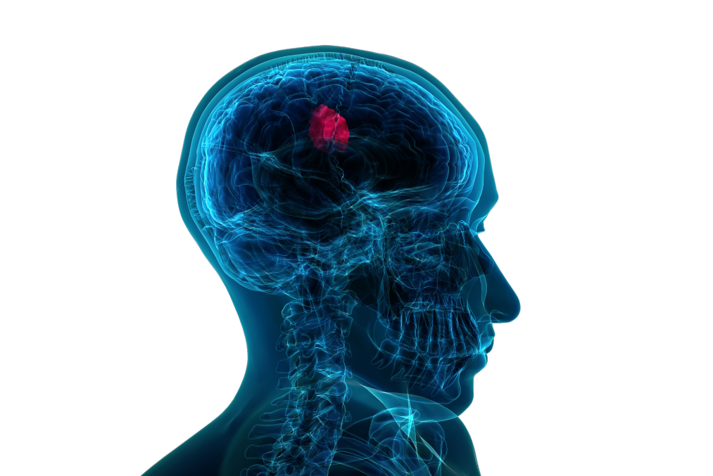


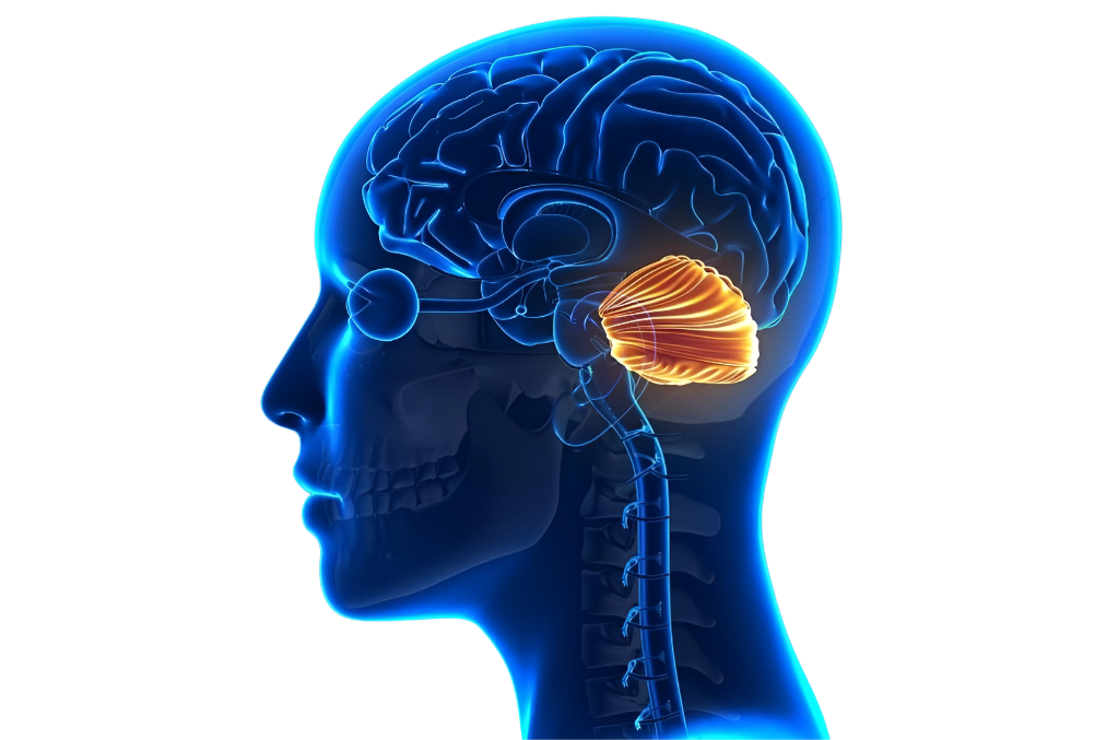
Some brain tumors do not cause symptoms for a long time or cause very mild complaints. Particularly, benign slow-growing masses and congenital tumors may remain undiagnosed for years and are sometimes discovered incidentally. General symptoms of brain tumors are well-defined: headaches, vomiting, visual impairment, double vision, seizures, weakness and numbness in the arms and legs, memory loss, speech difficulties, imbalance, walking disturbances, hormonal imbalances, and others.
PATIENT FAQ
Find answers to common questions.
A searchable FAQ for neurosurgery, treatment options, and patient guidance.
Search
39 questions
Sudden, severe, and persistent explosive headaches are significant and may indicate sudden bleeding inside the skull, requiring immediate examination. Some headaches, even if not severe, may be prolonged and dull in nature, suggesting the presence of a tumor inside the skull. However, most headaches are due to migraines, high blood pressure, or stress.
With advanced MRI techniques, benign and malignant tumors can now be distinguished with high accuracy. Additionally, auxiliary methods such as PET CT/MRI can be used. However, the exact nature of a tumor can only be definitively identified by pathological examination of its tissue.
Yes, it is possible to live with a brain tumor for years without experiencing any symptoms. Especially with slow-growing types of brain tumors, symptoms may not appear in the early stages.
No. For some tumors only surveillance is chosen, for others biopsy, and for others resection. The main deciding factors are anatomical accessibility and functional risk.
No. In some cases surveillance is appropriate, in others biopsy, and in others surgery is needed.
Every operation carries risk; the level depends on tumor location and proximity to critical brain functions.
The decision is based on tumor type, grade, location, size, the patient's neurological status, and the balance of surgical risk. Not every lesion must be removed directly.
Not always. Microscopic spread, tumor biology, and recurrence risk mean follow-up and sometimes adjuvant treatment continue. Complete resection does not always mean complete cure.
No. Infection, demyelinating disease, vascular lesions, and treatment-related changes can also appear as masses. MRI findings must be interpreted with clinical context and pathology when needed.
Not entirely. Most brain tumors are visible on MRI, but very small, early, or specially located lesions may need further evaluation. If clinical findings are strong, a single normal MRI does not close the workup.
First seizures in adulthood, focal seizure features, accompanying neurological deficits, and progressive symptoms increase tumor likelihood. Seizures are not always caused by tumors, but imaging is required.
Gradually worsening weakness that is not sudden, especially with headache, speech difficulty, or balance problems, suggests a central nervous system lesion. This is particularly important with cortical or deep-seated masses.
Speech problems may relate to the dominant hemisphere, especially frontal and temporal language areas. Whether aphasia or dysarthria predominates guides interpretation.
Glioblastoma is high-grade, aggressive, and fast-growing; low-grade gliomas may behave more slowly. Definitive distinction usually requires pathology and molecular testing.
Because of rapid growth, infiltration into surrounding tissue, and high recurrence risk. Treatment planning is usually multidisciplinary and time-sensitive.
Because residual disease and early recurrence risk exist. Follow-up MRI scans are needed to monitor treatment response and possible progression.
Primary tumors start in the brain; metastases are spread from cancer elsewhere. Distinction relies on clinical history, imaging pattern, and pathology when needed.
Yes. Behavioral and personality changes can occur, especially with frontal lobe involvement.
Yes. Children may have headache, vomiting, imbalance, developmental changes, and declining school performance.
Not always. Microscopic disease, tumor type, and recurrence risk may require continued follow-up or additional treatment.
Yes. Especially with some high-grade tumors, recurrence risk is significant.
When surgical decisions, radiation plans, pathology results, or treatment options are unclear, a second opinion is valuable.
No, not every incidental finding needs urgent surgery. Size, location, growth rate, age, and overall health are assessed together. Small asymptomatic low-risk lesions are often monitored with interval MRI; surgery is considered if growth or new symptoms appear. The decision depends on change over time, not a single scan.
Size alone is not decisive. Growth rate, pressure on surrounding tissue, age, general health, and expected quality of life are weighed together. Two tumors of the same size can lead to different decisions if only one compresses a critical area; there is no universal threshold for everyone.
Not always. The balance is between maximal tumor removal and preserving neurological function. Neuronavigation, intraoperative neuromonitoring, and awake craniotomy when needed help define limits in real time. Sometimes partial resection is chosen to protect function, with radiation or other therapies for residual disease.
That assessment depends on tumor location, the patient's condition at the time, and the center's imaging and surgical capabilities. Another center or multidisciplinary approach may change the outcome. Patients who receive such assessments, especially in complex cases, commonly seek a second opinion.
Brain and spine tumor care often has several reasonable paths among surgery, radiation, chemotherapy, and surveillance, so differing opinions are not unusual. Understand each recommendation's rationale (why this option, risks, and expected benefit) and, when possible, ensure discussion in a tumor board (oncologist, radiation oncologist, neurosurgeon).
Intraoperative neuromonitoring tracks electrical responses of nerve pathways throughout surgery and alerts the surgeon early. For tumors very close to speech or motor areas, awake craniotomy with direct speech/movement testing may be used. These methods do not eliminate risk entirely but allow much finer functional mapping.
Generally yes — repeat surgery can be more complex because scar tissue and altered anatomy from the first operation complicate planning. The degree of added risk depends on location, time since the first surgery, and prior radiation. Reoperation decisions are again individualized through multidisciplinary review.
In most cases, a specific cause cannot be identified. However, epilepsy can result from conditions such as head trauma, stroke, lack of oxygen to the brain, or brain infections.
An electroencephalogram (EEG) test is performed to detect abnormal electrical activity in the brain, which is typical in epilepsy. In this test, small sensors are attached to the scalp to capture the electrical signals generated when brain cells communicate with each other, allowing for the detection of abnormalities.
Approximately 70% of epilepsy patients respond to medication. In the remaining cases, seizure frequency is reduced. However, about 25% of patients do not respond adequately to medication, which is referred to as drug-resistant epilepsy.
For drug-resistant and suitable patients, surgical treatment is an option. However, surgery is not applicable for all patients and requires thorough testing to determine eligibility. Surgical treatment does not always completely eliminate seizures and may not benefit some patients or could potentially worsen their condition. Surgical options include removing the group of cells causing seizures or preventing the spread of electrical signals to other areas. Additional techniques include placing electrodes in specific areas of the brain (deep brain stimulation) or vagus nerve stimulation.
Rarely yes, especially in genetic syndromes such as neurofibromatosis type 2, where multiple tumors may develop along the brain and spine. Treatment plans consider all tumors and usually prioritize the one posing the greatest functional risk. Genetic counseling may be included when such a history exists.
Spinal tumors are often metastatic tumors originating from another type of cancer. Treatment responses can vary depending on the patient's condition. While brain tumors with rapid progression may lead to fatal outcomes, spinal cord tumors differ in this respect. If a spinal cord tumor is located near critical areas such as the brain, heart, or respiratory center, there may be a risk of death. Otherwise, spinal cord tumors may not cause death but can lead to paralysis over time if left untreated. A patient with a spinal cord tumor can continue living, but without proper treatment, they face risks such as losing the ability to use their limbs or paralysis below the tumor's location.
Tumors developing within the spinal cord typically do not metastasize. However, certain childhood brain tumors located along the circulation pathway of cerebrospinal fluid may migrate toward the spinal cord. Primary spinal cord tumors themselves generally do not metastasize.
Spinal and spinal cord tumors can indeed cause paralysis. Untreated or late-treated tumors may lead to paralysis over time or suddenly. Paralysis caused by these tumors depends on their location and can affect the body below the neck or waist.
Recovery after spinal and spinal cord tumor surgeries varies depending on the type of tumor, its location, and the patient's response to treatment. Generally, patients can return to their social lives about three weeks after surgery.
No matching questions.