

Neuromonitoring
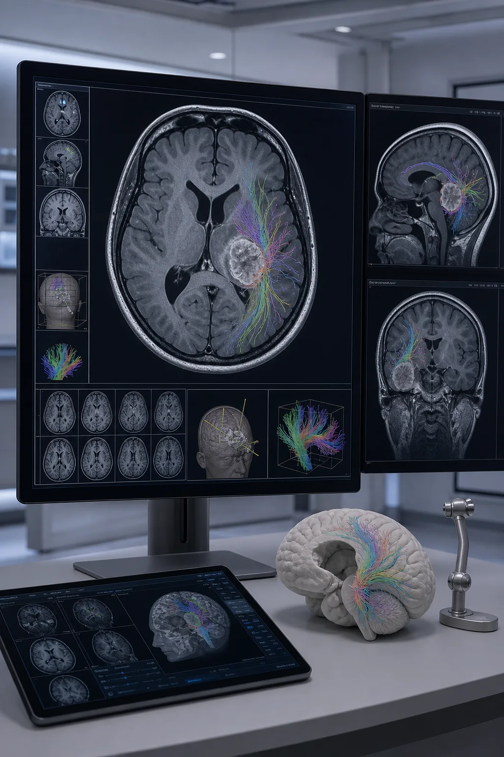
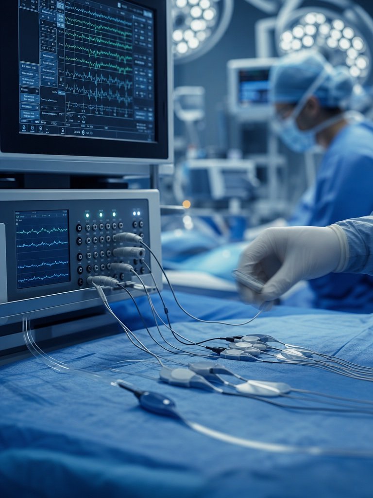
Real-time monitoring of nervous system functions during surgery, used to minimize complications and improve surgical outcomes.


Accurate Diagnosis and Effective Treatment
Spinal Cord Tumor
Spinal cord tumors can be controlled with early diagnosis and proper treatment. Let your pain and movement restrictions end; take a step towards a healthy life.
Share details and at least one file.
Assoc. Prof. Dr. Güçlühan Güçlü is a neurosurgery specialist.
His clinical focus includes neuro-oncology, functional brain surgery and spine surgery; assessment is individualized, and treatment plans are set after examination.
He earned his medical degree at Dokuz Eylül University Faculty of Medicine (1990–1996) and completed his neurosurgery residency at Bakırköy Prof. Dr. Mazhar Osman Mental Health and Neurological Diseases Training and Research Hospital (1997–2003). From 2003 to 2023 he served as a neurosurgery specialist / associate professor at Bakırköy Dr. Sadi Konuk Training and Research Hospital, and as Clinical Administrative Officer from 2018 to 2023.
Diagnosis and Treatment
Real-time monitoring of nervous system functions during surgery, used to minimize complications and improve surgical outcomes.
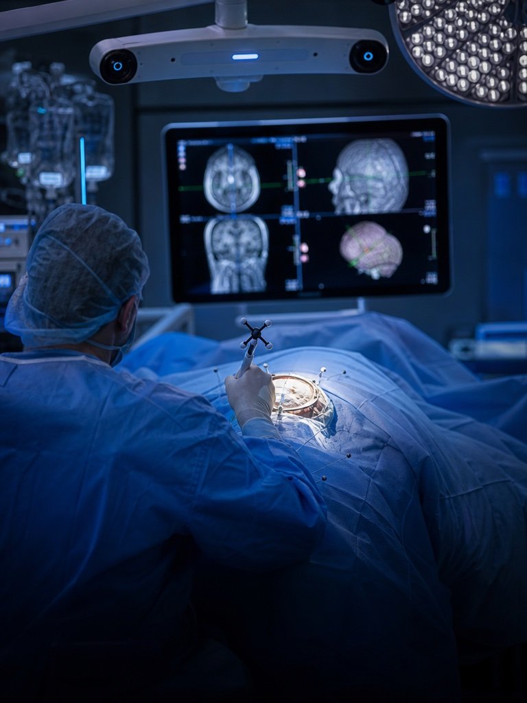
Advanced imaging technology that assists the surgeon in accurately identifying tumors and other target structures during brain surgery.
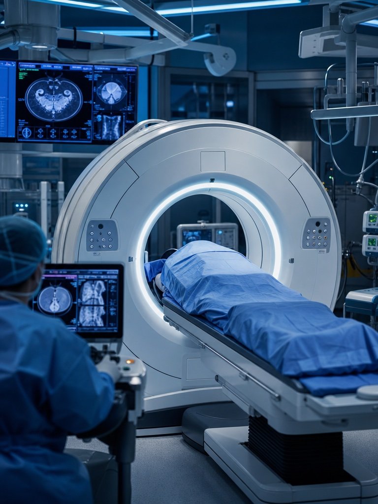
Enhances the accuracy of surgical interventions by providing real-time computed tomography (CT) images during surgery.
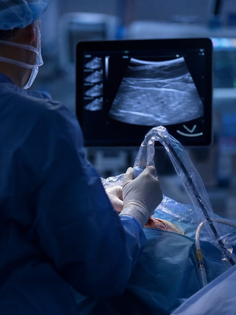
Assists in identifying tumors and other abnormalities during surgery using ultrasound imaging.
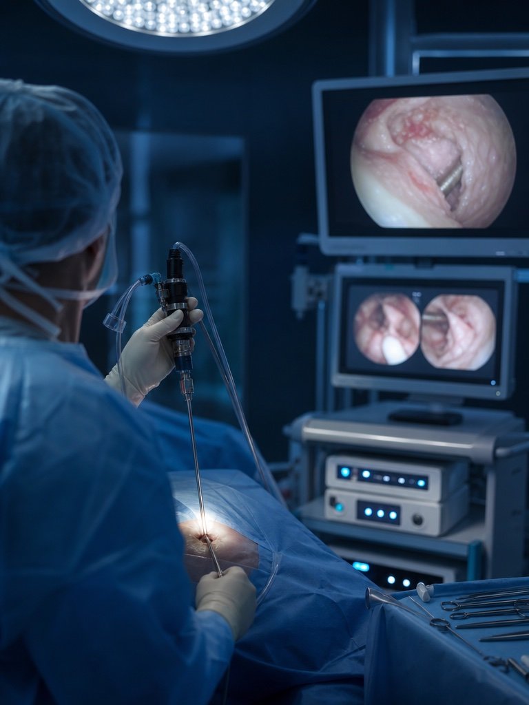
Utilizes minimally invasive surgical techniques to perform brain and spinal surgeries through small incisions with the help of an endoscope.
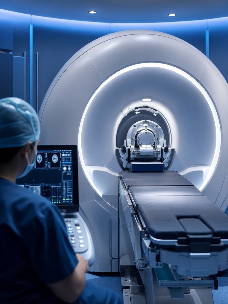
A device used to precisely deliver high doses of radiation to treat brain tumors and other brain abnormalities.
Blog
Read the latest blog posts by Assoc. Prof. Dr. Güçlühan Güçlü on current developments and healthy living.
FAQ
Learn About Neurosurgery...
Book appointmentSome brain tumors do not cause symptoms for a long time or cause very mild complaints. Particularly, benign slow-growing masses and congenital tumors may remain undiagnosed for years and are sometimes discovered incidentally. General symptoms of brain tumors are well-defined: headaches, vomiting, visual impairment, double vision, seizures, weakness and numbness in the arms and legs, memory loss, speech difficulties, imbalance, walking disturbances, hormonal imbalances, and others.
Sudden, severe, and persistent explosive headaches are significant and may indicate sudden bleeding inside the skull, requiring immediate examination. Some headaches, even if not severe, may be prolonged and dull in nature, suggesting the presence of a tumor inside the skull. However, most headaches are due to migraines, high blood pressure, or stress.
With advanced MRI techniques, benign and malignant tumors can now be distinguished with high accuracy. Additionally, auxiliary methods such as PET CT/MRI can be used. However, the exact nature of a tumor can only be definitively identified by pathological examination of its tissue.
Yes, it is possible to live with a brain tumor for years without experiencing any symptoms. Especially with slow-growing types of brain tumors, symptoms may not appear in the early stages.
No. For some tumors only surveillance is chosen, for others biopsy, and for others resection. The main deciding factors are anatomical accessibility and functional risk.
No. In some cases surveillance is appropriate, in others biopsy, and in others surgery is needed.
Every operation carries risk; the level depends on tumor location and proximity to critical brain functions.
The decision is based on tumor type, grade, location, size, the patient's neurological status, and the balance of surgical risk. Not every lesion must be removed directly.
Not always. Microscopic spread, tumor biology, and recurrence risk mean follow-up and sometimes adjuvant treatment continue. Complete resection does not always mean complete cure.
No. Infection, demyelinating disease, vascular lesions, and treatment-related changes can also appear as masses. MRI findings must be interpreted with clinical context and pathology when needed.
Not entirely. Most brain tumors are visible on MRI, but very small, early, or specially located lesions may need further evaluation. If clinical findings are strong, a single normal MRI does not close the workup.
First seizures in adulthood, focal seizure features, accompanying neurological deficits, and progressive symptoms increase tumor likelihood. Seizures are not always caused by tumors, but imaging is required.
No matching questions.