



At a glance
No, finding a tumor in the brain does not always mean that surgery is needed. For some patients, regular clinical and imaging follow-up may be sufficient; for others, a biopsy to establish a definitive diagnosis or removal of the tumor may be considered. The decision about brain tumor surgery is based not only on MRI images, but on a combined assessment of the likely tumor type, location, growth behavior, symptoms, pressure on surrounding tissues, whether it can be removed safely, and the patient's overall condition. The decision frameworks for meningioma, diffuse glioma or glioblastoma, and brain metastases differ; the shared question is whether the expected diagnostic or therapeutic benefit outweighs the risk of losing neurological function. [1–5]
Does every brain tumor require surgery?
Not every brain tumor requires surgery. “Brain tumor” describes not a single disease, but a broad range of lesions with different biological behaviors, growth patterns, and treatment approaches.
A selected meningioma found incidentally and causing no symptoms may be followed with active monitoring. [2,7] In diffuse gliomas, tissue diagnosis and molecular characteristics may determine the treatment plan. [3] In selected patients with newly diagnosed supratentorial glioblastoma, surgery to reduce the tumor burden as safely as possible (cytoreductive surgery) is considered; other illnesses, functional status, and tumor location influence the choice between biopsy, subtotal resection, and more extensive resection. [5] For brain metastases, the number and size of tumors, mass effect, the status of the systemic cancer, and other local treatment options are assessed together. [4]
There is no single tumor size, age limit, or MRI criterion that applies to everyone. Imaging is a key part of the decision, but it cannot, on its own, establish the exact tumor type, the patient's functional reserve, or the expected benefit of an intervention. The surgical decision is shaped by the images, neurological examination, progression of symptoms, previous investigations, and guidelines specific to the tumor group, with multidisciplinary assessment when needed. [1–5]
What factors are assessed when deciding on brain tumor surgery?
The decision depends on interpreting interrelated clinical, imaging, and functional factors together, rather than on a single finding. Two tumors of the same size may lead to different options in different locations and different patients.
Tumor type and biological behavior
The approach differs depending on whether the tumor is a slow-growing, well-defined lesion, an infiltrative tumor extending into surrounding brain tissue, or a cancer that has spread to the brain from another organ. In some cases, imaging strongly suggests a provisional diagnosis; however, tissue examination is needed for definitive classification and, particularly in gliomas, for molecular characteristics that affect treatment. [2–4]
Tumor location in the brain
Areas involved in language, movement, vision, memory, and cognitive functions, as well as proximity to the brainstem, deep nuclei, major blood vessels, and important nerve pathways, influence surgical planning. A difficult location does not automatically mean that surgery is impossible, but it changes the intended extent of removal, the method used, and the acceptable risk. [1,6]
Growth rate and previous imaging
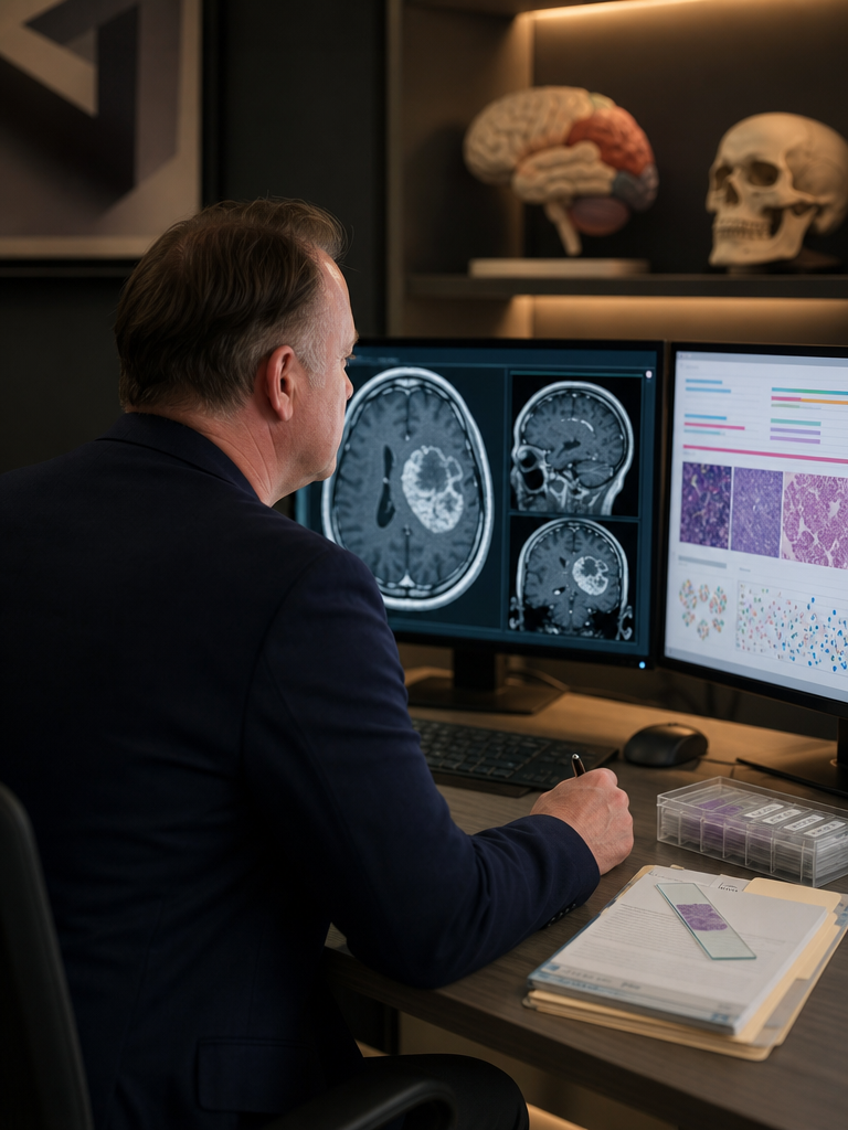
A single MRI may not show how a tumor behaves over time. Comparison with previous images helps assess whether the lesion has remained stable, its growth rate, edema, and its effects on surrounding structures. Serial MRI is particularly important for assessing behavior over time in cases where imaging suggests a meningioma and active monitoring is planned. Growth alone does not automatically lead to a decision to operate; it is interpreted alongside other clinical and imaging findings. [2,7]
Symptoms and neurological examination findings
Progressive weakness, language difficulties, visual problems, cognitive changes, or seizures that are difficult to control are important in surgical assessment. Whether the symptoms are actually caused by the tumor and how likely they are to improve after pressure is relieved are assessed separately. [1,4]
Mass effect, edema, and hydrocephalus
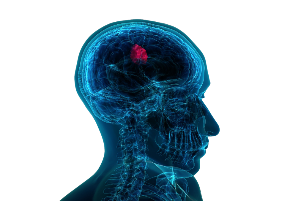
The displacement of surrounding tissues by a tumor is called “mass effect,” an increase in fluid around it is called “edema,” and disruption of cerebrospinal fluid circulation is called “hydrocephalus.” These conditions may increase pressure inside the skull and raise the need for priority intervention. The brain metastases guideline also states that large tumors causing mass effect may be more likely to benefit from surgery. [4]
The patient's age, general health, and functional status
Age alone is not an automatic criterion for surgery or for monitoring without surgery. Particularly when assessing brain metastases and newly diagnosed supratentorial glioblastoma, heart and lung disease, medications, frailty, independence in daily life, existing neurological deficits, functional status, and coexisting systemic illnesses affect the expected benefit and the likelihood of tolerating the intervention. [4,5]
Safe resectability and expected neurological outcome
The goal of surgery is not simply to remove as much tissue as possible, but to do so while preserving function. The concept of maximal safe resection, used particularly in glioma surgery, means removing as much tumor tissue as possible up to the functional boundaries that protect language, movement, vision, cognition, memory, and independent living. [1]
Beyond this limit, leaving less tumor visible on imaging may not mean a better outcome for the patient. The current glioma surgery guideline, drawing largely on observational and prognostic data, emphasizes that the extent of resection should be assessed alongside neurological status, neurocognitive functions, seizures, quality of life, and the planning of subsequent treatments, as well as oncological outcomes. In meningioma surgery, too, a fundamental principle is that neurological or cognitive function should not be sacrificed for more extensive removal. [1,2]
When might active monitoring be considered?
Active monitoring is a planned and supervised approach for selected patients; it does not mean “doing nothing.” It may be considered particularly in selected cases with imaging features consistent with meningioma, found incidentally and causing no symptoms, if there is no significant mass effect or hydrocephalus. The EANO meningioma guideline considers this an accepted option; prospective meningioma cohorts provide information about the natural course but do not, on their own, determine an individual threshold for intervention. [2,7]
The follow-up plan should include MRI intervals, clinical reviews, and the changes that would require reassessment. During meningioma monitoring, new symptoms, changes in imaging behavior, or increasing effects on surrounding structures may require the plan to be reconsidered. [2]
The monitoring interval is not the same for every tumor. It is determined by the likely tumor type, imaging characteristics, and the patient's age and overall condition. For example, a monitoring approach used for benign tumors suspected to be meningiomas on imaging cannot routinely be applied to diffuse gliomas or brain metastases; these groups have different diagnostic and treatment pathways. [2–4]
When might a brain tumor biopsy be preferred?
A biopsy may be preferred, particularly when a diffuse glioma or another infiltrative tumor is suspected, if a histopathological and molecular diagnosis is needed to guide treatment but the expected benefit of extensive surgical removal is lower than the risk to function. The aim is not to remove the whole tumor, but to obtain an adequate, suitable tissue sample for diagnosis. [3,5]
If a lesion suspected to be a diffuse glioma or infiltrative tumor is deep-seated, located in the midline, multifocal, or close to areas responsible for critical functions, and safe, clinically meaningful resection does not appear possible, stereotactic biopsy may provide a diagnosis through a more limited intervention. However, biopsy is not risk-free and is not an appropriate option for every lesion. [3,5]
Surgical resection may aim to reduce the tumor burden, relieve pressure, or achieve local control, in addition to providing a diagnosis. Biopsy and resection should therefore not be viewed as the same procedure, or simply smaller and larger versions of one another. The guideline for newly diagnosed supratentorial glioblastoma recommends choosing between biopsy, subtotal resection, and more extensive resection by considering tumor location, the patient's functional status, and other illnesses. [5]
When is brain tumor surgery needed?
Surgery is considered when there is a possibility of reducing the tumor's current or anticipated near-term harm, establishing a diagnosis, relieving pressure, or achieving a clinically meaningful reduction in tumor burden. Findings such as progressive neurological deficits, marked mass effect, increased pressure inside the skull, hydrocephalus, difficult-to-control seizures, or growth on imaging may carry weight in the assessment; however, they do not form a single list of surgical indications applicable to all tumors. Symptoms and growth in meningioma, the need for safe cytoreduction and tissue diagnosis in gliomas, and selected brain metastases that are particularly large and cause mass effect are assessed differently. [1–5]
It is also important that the tumor is accessible, that a safe surgical corridor is available, and that clinically meaningful removal can be achieved within functional limits. The need for surgery is not assessed by the same criteria for meningioma, diffuse glioma or glioblastoma, and brain metastases. [1–5]
The purpose of surgery may vary according to the tumor type and the patient's condition: a definitive tissue diagnosis, relief of pressure, reduction of seizure burden, reduction of tumor volume, or local control may be one or more of its goals. [1–5]
Is removing the entire tumor always the right goal?
No. Surgical success cannot be defined solely by the absence of residual tumor on postoperative MRI. Preserving neurological functions, cognitive capacity, and the person's ability to live independently is an integral part of the treatment outcome.
While glioma guidelines report an association between the extent of resection and oncological outcomes, largely based on observational and prognostic data, they also emphasize that new neurological deficits may affect quality of life and subsequent treatments. The goal is therefore not to remove the tumor seen on imaging at any cost, but to achieve clinically meaningful surgical benefit without causing an unacceptable loss of function. [1]
When the tumor approaches networks involved in language, movement, vision, or memory, the surgeon may reach a predefined functional stopping boundary. In this situation, deliberately leaving part of the tumor behind may be a conscious choice to avoid the risk of permanent loss of function from further removal.
This does not mean that the operation has failed. In glioma and meningioma surgery, oncological benefit must be balanced against outcomes that matter to the patient, such as language, movement, cognitive function, and independent living. [1,2]
Monitoring, radiosurgery, fractionated radiotherapy, systemic treatment, or combinations of these may be considered for residual tumor tissue. The appropriate additional treatment depends on the tumor type, such as meningioma, diffuse glioma, glioblastoma, or metastasis, and the pathological and molecular findings. [2–4]
The approach to tumors near functional brain areas
A tumor close to a functional area is not automatically “inoperable.” Particularly in gliomas, this location may require more detailed surgical planning and removal guided by functional boundaries as well as anatomical imaging. [1]
Depending on the tumor type, location, the patient's condition, and the resources available at the center, the following methods may be used during planning or surgery:
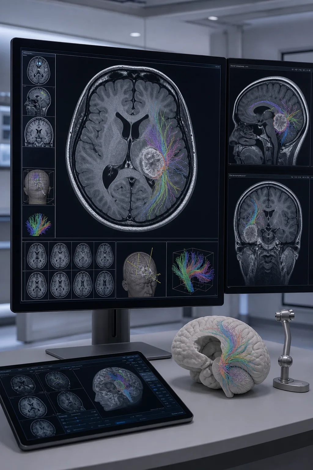
- Functional MRI: Helps assess the approximate locations of brain areas involved in specific tasks before surgery.
- Neuronavigation: Allows MRI images to be used for anatomical guidance during surgery.
- Neuromonitoring: Helps monitor signals associated with nerve pathways and neurological functions during the operation.
- Awake surgery: May allow language, movement, or other selected functions to be tested during the operation in suitable patients.
- Cortical and subcortical mapping: Aims to assess functional boundaries on the brain surface and in deeper areas using electrical stimulation.
A single-center prospective observational series of selected patients with supratentorial gliomas near functional areas reported that resection guided by functional boundaries could be performed using awake cortical and subcortical mapping. This study, which had no control group, does not establish cause and effect, and its results cannot be generalized to every patient or tumor type. [6]
None of these methods alone guarantees freedom from risk or complete removal. Their purpose is to try to reduce the likelihood of functional loss by defining the surgical corridor and stopping boundaries more precisely.
Why does the decision differ by tumor type?
The same size on imaging does not mean the same thing for different tumors. The table below is not a personal treatment recommendation, but a brief summary of the main differences in decision-making.
| Tumor group | Main factors affecting the decision | Possible approach |
|---|---|---|
| Meningioma | Symptoms, growth, edema, location, and proximity to critical structures | Active monitoring in selected incidental, asymptomatic cases; surgery for growing or symptomatic tumors; radiosurgery or radiotherapy in some cases [2,7] |
| Diffuse glioma and glioblastoma | Infiltrative growth, histomolecular diagnosis, safe resectability, and neurological functions | Tissue diagnosis is important; maximal safe resection when it can be performed safely, or biopsy if it is not appropriate; followed by additional treatments according to tumor type [1,3,5] |
| Brain metastases | Number and size of lesions, mass effect, symptoms, systemic disease, and performance status | Surgery for selected large lesions causing pressure; a multidisciplinary plan involving radiosurgery, radiotherapy, and systemic treatments [4] |
A deep or critical location is not, in itself, a treatment category. The same anatomical feature may affect different choices: selected monitoring, planned limited removal, or radiotherapy in meningioma; stereotactic biopsy or resection guided by functional boundaries in diffuse glioma; and the balance between surgery, radiosurgery, and systemic treatment in brain metastases. [1–4]
How are the risks of brain tumor surgery assessed?
Risk cannot be expressed as a single rate for all brain tumor operations. The tumor type and location, its relationship to blood vessels and nerve pathways, the purpose of surgery, the intended extent of removal, previous treatments, and the patient's overall condition change the risk profile. The items below are possible risks that vary by context, not outcomes expected at the same frequency in every patient. [1,2,5,6]
- Temporary or permanent weakness
- Difficulty understanding or expressing language
- Effects on vision, balance, memory, or cognitive functions
- Seizures
- Bleeding
- Infection
- Brain edema
- Problems with cerebrospinal fluid circulation
- General complications related to anesthesia
Not every risk has the same likelihood in every patient. Rates found online cannot be translated into a personal risk estimate without knowing the tumor type, location, surgical goal, extent of the procedure, and patient group. [1,2,5,6]
The risk–benefit assessment includes not only the risk of surgery, but also the risk that the particular tumor type may pose without intervention. The potential neurological and functional cost of achieving the expected surgical benefit must be assessed clearly. [1,2,4–6]
When might a second opinion on a brain tumor be helpful?
A second opinion may be helpful when the diagnosis or tumor type is uncertain, or when there is more than one reasonable option among active monitoring, biopsy, and surgery.
- The tumor is in a critical area of the brain
- A decision is needed between biopsy and resection
- There are different approaches to the extent of surgery
- It is uncertain whether the entire tumor can be removed safely
- Radiosurgery, radiotherapy, and surgery have been recommended in different sequences
- The pathology or molecular diagnosis needs to be reassessed
The purpose of a second opinion is not to dismiss the first recommendation, but to reassess the imaging, likely diagnosis, expected extent of removal, functional risks, and subsequent treatment plan with another specialist team.
Bringing current and previous MRI images, radiology reports, any pathology results, a list of medications, and information about the progression of symptoms helps the assessment.
Conclusion
The surgical decision depends not simply on the presence of a tumor, but on its behavior, the risk it poses to the patient, and whether safe intervention is possible. Active monitoring, biopsy, and surgical resection are options that serve different clinical purposes.
The appropriate approach considers the likely tumor type, location and changes over time, the patient's symptoms, overall condition, and priorities together. The same decision algorithm is not used for meningioma, diffuse glioma or glioblastoma, and brain metastases. The aim is not simply to reduce the amount of tumor, but to balance diagnostic and oncological benefit in the context of the particular disease with neurological functions and quality of life. [1–5]
Frequently asked questions
Can a benign brain tumor be operated on?
Yes, some benign brain tumors can be operated on; however, this description alone does not mean that surgery is needed or that the lesion is insignificant. Symptoms, growth, edema, pressure, location, and whether the tumor can be removed safely are assessed together. Active monitoring may be appropriate for selected asymptomatic meningiomas. [2,7]
Does a small brain tumor require surgery?
Small size alone is not a sufficient basis for a decision. A small lesion in a critical area can cause significant symptoms, while a larger lesion elsewhere may be monitored in selected circumstances. Tumor type, growth behavior, and its effect on the patient are more decisive.
Can a brain tumor be definitively diagnosed by MRI alone?
MRI provides valuable information about the location, size, and imaging characteristics of a lesion and may strongly suggest a provisional diagnosis for some tumors. However, definitive classification, particularly of molecular characteristics, usually relies on tissue examination. The need for biopsy or resection is determined by the clinical context. [2,3]
What is the difference between a biopsy and brain tumor surgery?
The main purpose of a biopsy is to obtain a limited amount of tissue for diagnosis. Surgical resection may aim to reduce tumor volume, relieve pressure, or achieve local control, in addition to providing a diagnosis. The appropriate procedure is assessed according to the expected benefit and risk to function. [3,5]
What happens if the tumor cannot be completely removed?
The significance of remaining tissue depends on the tumor type. Monitoring, further surgery, radiosurgery, radiotherapy, or systemic treatment may be planned according to the pathological and molecular findings and the patient's condition. Deliberately leaving some tumor behind to preserve function may be the appropriate surgical limit for some patients. [1–4]
Can older people undergo brain tumor surgery?
Older age alone does not automatically rule out surgery. Particularly in selected patients with newly diagnosed glioblastoma, performance status, frailty, other illnesses, tumor location, and the surgical goal are assessed together; much of the evidence comes from observational data on patients selected as candidates for surgery. For other tumor types, decisions are also specific to the patient and the disease. [5]
Can a brain tumor be treated without surgery?
Is the decision to operate based only on MRI findings?
No. Although MRI is very important, the neurological examination, progression of symptoms, previous imaging, general health, functional status, likely pathology, and whether the tumor can be removed safely also contribute to the decision. A multidisciplinary assessment is carried out when needed. [1–5]
References
- 1. Goldbrunner R, Foroglou N, Signorelli F, et al. EANS–EANO Guidelines on the Extent of Resection in Gliomas. Neuro-Oncology. 2026;28(1):38–54. doi:10.1093/neuonc/noaf217. doi:10.1093/neuonc/noaf217
- 2. Goldbrunner R, Stavrinou P, Jenkinson M, et al. EANO Guideline on the Diagnosis and Management of Meningiomas. Neuro-Oncology. 2021;23(11):1821–1834. doi:10.1093/neuonc/noab150. doi:10.1093/neuonc/noab150
- 3. Weller M, van den Bent M, Preusser M, et al. EANO Guidelines on the Diagnosis and Treatment of Diffuse Gliomas of Adulthood. Nature Reviews Clinical Oncology. 2021;18:170–186. doi:10.1038/s41571-020-00447-z. doi:10.1038/s41571-020-00447-z
- 4. Vogelbaum MA, Brown PD, Messersmith H, et al. Treatment for Brain Metastases: ASCO–SNO–ASTRO Guideline. Journal of Clinical Oncology. 2022;40(5):492–516. doi:10.1200/JCO.21.02314. doi:10.1200/JCO.21.02314
- 5. Domino JS, Ormond DR, Germano IM, et al. Cytoreductive Surgery in the Management of Newly Diagnosed Glioblastoma in Adults. Journal of Neuro-Oncology. 2020;150:121–142. doi:10.1007/s11060-020-03606-5. doi:10.1007/s11060-020-03606-5
- 6. Pallud J, Dezamis E. Functional and Oncological Outcomes Following Awake Surgical Resection Using Intraoperative Cortico-Subcortical Functional Mapping for Supratentorial Gliomas Located in Eloquent Areas. Neurochirurgie. 2017;63(3):208–218. doi:10.1016/j.neuchi.2016.08.003. doi:10.1016/j.neuchi.2016.08.003
- 7. Delgado-López PD, Montalvo-Afonso A, Martín-Alonso J, et al. Volumetric Growth Rate of Incidental Asymptomatic Meningiomas: A Single-Center Prospective Cohort Study. Acta Neurochirurgica. 2021;163(6):1665–1675. doi:10.1007/s00701-021-04815-1. doi:10.1007/s00701-021-04815-1