



What are the symptoms of a brain tumour?
Brain tumours can present with a wide range of symptoms, including headache, nausea and vomiting, seizures, weakness or numbness in an arm or leg, changes in speech or vision, balance problems, cognitive slowing and personality changes. However, none of these symptoms alone establishes a diagnosis of a brain tumour. Symptoms such as headache, dizziness and forgetfulness have much more common causes in the general population.[3–6]
What raises concern is often not the presence of a single symptom, but whether it is new, progressively worsening, different from the person's usual pattern, or accompanied by other neurological signs. Diagnosis relies on a detailed history, neurological examination, brain imaging when needed and, if a mass is found, sometimes tissue and molecular analysis.[1,2,7–10]
Why do brain tumours cause different symptoms?
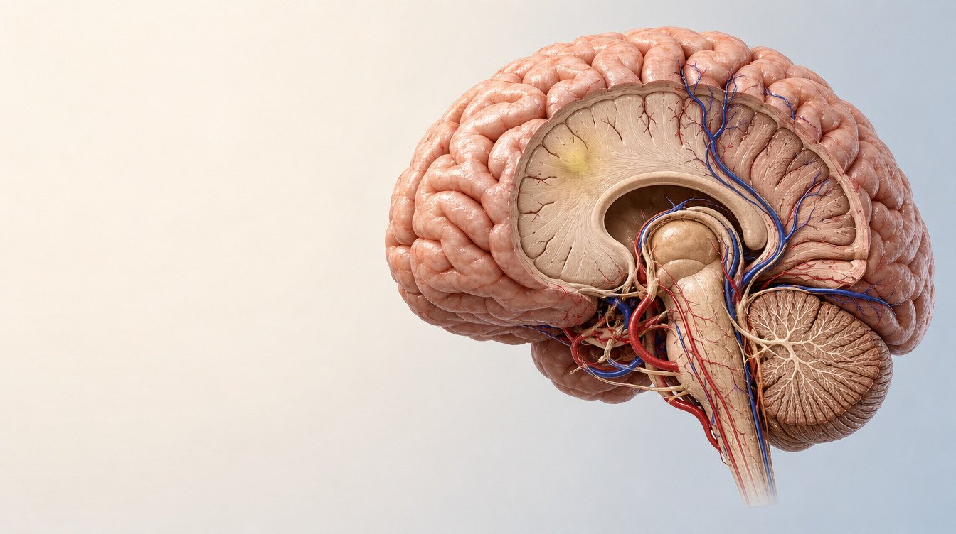
“Brain tumour” is not the name of a single disease. There are primary tumours arising from the brain or surrounding tissues, metastases that spread to the brain from another part of the body, and numerous subtypes occurring in different age groups. The current World Health Organization classification considers both microscopic appearance and molecular features for many central nervous system tumours.[8]
Symptoms may vary according to:
- Which part of the brain the tumour is in
- Its size and growth rate
- Whether it causes swelling around it
- Whether it obstructs the circulation of cerebrospinal fluid
- Whether it raises intracranial pressure
- Whether it disrupts the brain's electrical activity and causes seizures
- The person's age, general health and coexisting conditions
For this reason, a small lesion in a functionally critical area may cause marked signs, while another, slow-growing lesion may cause few symptoms for a long time. Symptom severity alone also does not show whether a tumour is “benign” or “malignant”.[6–10]
Main symptom patterns that may occur in adults

New or changing headache
Headaches are common, and the vast majority of people with headaches do not have a brain tumour. A study of a particular group of patients urgently referred with suspected brain cancer reported that, although headache was the most common reason for referral, most final diagnoses were migraine and isolated headache was not a reliable indicator of brain cancer.[4] This study involved a selected referral population; its rates should not be applied to everyone in the general population.
Headache features that lower the threshold for assessment include:
- A new headache that has not occurred before
- Increasing frequency or severity over days or weeks
- A marked difference from the person's established migraine or headache pattern
- Accompanying seizures, weakness, numbness or changes in speech, vision or balance
- Worsening with coughing, straining or exercise
- A marked change with position
- Additional risks such as a history of cancer, immunosuppression, fever or papilloedema
These features do not mean “there is a tumour”. In the SNNOOP10 approach, these and similar red flags prompt investigation for secondary causes of headache, including bleeding, infection, vascular disease and disorders of intracranial pressure, as well as tumours.[3]
Nausea and repeated vomiting
Nausea and vomiting can have many causes, including migraine, infection, pregnancy, medication effects and digestive disorders. However, if a headache is accompanied by repeated vomiting, blurred vision, drowsiness, marked imbalance or altered consciousness, causes such as raised intracranial pressure need to be ruled out promptly.[5]
The idea that “morning vomiting is a definite sign of a brain tumour” is incorrect. Headache and vomiting that are more pronounced in the morning can occur with raised intracranial pressure, but this pattern is neither present in every patient nor specific to a tumour.[5]
A first seizure

A first seizure in adulthood may be associated with a metabolic disorder, vascular disease, infection, trauma or other causes besides a brain tumour. Nevertheless, a first seizure requires medical assessment without delay.[11,12]
A seizure does not always involve whole-body convulsions. Focal seizures may involve brief staring episodes, unresponsiveness, rhythmic movements of one arm or leg, repeated purposeless behaviour, an unusual smell or taste, or a brief speech disturbance. Call 112 if a seizure lasts more than five minutes, recurs before the person has fully regained consciousness, or is accompanied by breathing difficulty or serious injury. During a seizure, the person should not be restrained, nothing should be put in their mouth, and they should be turned safely onto their side.[12]
Weakness, numbness and speech disturbance

Facial asymmetry, new weakness in one arm or leg, numbness on one side, difficulty finding words or understanding speech are focal neurological symptoms. They can occur with a brain tumour, but can also be signs of much more time-sensitive conditions such as stroke or bleeding in the brain. Particularly when symptoms start suddenly, the person should seek emergency help rather than trying to distinguish the cause themselves.[1,11]
One-sided weakness or loss of central neurological function that progresses over hours, days or a few weeks may also require prompt imaging and specialist assessment. NICE recommends considering urgent brain MRI, or CT if MRI is unsuitable, in adults with progressive, subacute loss of central neurological function.[1]
Changes in vision, hearing, balance and walking
Loss of part of the visual field, double vision, new marked blurring, changes in eye movements, hearing loss on one side, imbalance or poor coordination can occur depending on the lesion's location. However, eye, inner ear, vascular and other neurological disorders can cause the same symptoms.
New loss of vision, marked difficulty walking or rapidly worsening imbalance should not be left unassessed, particularly when accompanied by other neurological signs. Finding papilloedema, swelling of the optic disc, during examination of the back of the eye suggests raised intracranial pressure and requires prompt investigation.[3,5]
Forgetfulness, cognitive slowing and personality changes
Much more common causes of forgetfulness include sleep deprivation, depression, stress, medications, thyroid and vitamin disorders, and age-related changes. Forgetfulness alone does not establish a diagnosis of a brain tumour.
By contrast, difficulty planning, slowed speech, a marked decline in work or everyday skills, impaired impulse control, or changes in personality and behaviour noticed by relatives that develop over a short period and progress require neurological assessment. The patient may not recognise the change, making a relative's observations valuable. Although symptoms can be related to location, a lesion's site or type cannot be determined from behavioural changes alone.[4,6,17]
Hormonal changes
Lesions in or around the pituitary gland may cause irregular periods, changes in sexual function, milk discharge from the breasts, altered growth, excessive thirst and urination, or visual field loss. These symptoms can also occur in many hormonal disorders. Assessment often requires collaboration between endocrinology, ophthalmology, neurology and, when needed, neurosurgery.
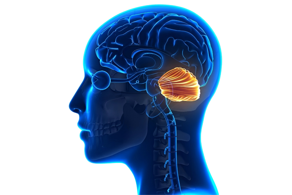
Which symptoms may be associated with different parts of the brain?
Certain areas of the brain are more closely associated with particular functions. This knowledge helps clinicians interpret examination findings, but does not allow someone to predict a tumour's location from their own symptoms. Swelling, mass effect, connections between brain networks and the person's dominant cerebral hemisphere can alter the clinical picture.[6,9,17]

| Region | Possible findings | Why is this not diagnostic? |
|---|---|---|
| Frontal lobe | Difficulty planning and making decisions, changes in personality and behaviour, weakness on the opposite side, difficulty producing speech | Vascular diseases, psychiatric conditions, medications and neurodegenerative diseases can cause similar findings |
| Temporal lobe | Focal seizures, memory changes, problems with understanding language or hearing | These can overlap with migraine, epilepsy and other neurological diseases |
| Parietal lobe | Numbness, impaired spatial perception, difficulty recognising one side of the body, problems with calculation or orientation | The distribution of symptoms does not precisely delineate a lesion's boundaries |
| Occipital lobe | Visual field loss, changes in the perception of light or shapes | These can be confused with symptoms originating in the eye, retina or optic nerve, or from migraine |
| Cerebellum | Problems with balance, coordination, fine movements and walking | Inner ear disorders, medications, alcohol, vascular and metabolic causes may be more common |
| Brainstem | Double vision, difficulty swallowing or speaking, eye movement abnormalities or signs involving several cranial nerves | Brainstem symptoms can also occur in time-sensitive vascular and inflammatory diseases |
| Pituitary gland and surrounding area | Visual field loss, changes in menstruation or sexual function, growth or fluid balance disturbances | Primary hormonal disorders can cause the same symptoms |
For example, one-sided weakness can occur with a brain lesion affecting the motor pathways, but when it starts suddenly, emergency causes such as stroke are considered first. Similarly, visual flashes may be associated with migraine aura or a focal seizure. Clinical decisions are based on the duration and manner of onset of symptoms, together with examination and imaging.
Do symptoms mean there is a tumour, and does their absence rule one out?
No. Most common symptoms have causes other than a tumour. Conversely, some brain tumours are found incidentally on imaging performed for another reason. Ticking many items on an online symptom list therefore does not reliably calculate the likelihood of a diagnosis, and ticking none does not provide absolute reassurance.
A mass or another lesion seen on imaging does not, by itself, mean “cancer”. Some lesions, such as meningiomas, may grow slowly; vascular structures, infections, inflammatory diseases and treatment-related changes can also mimic a mass on imaging. Radiological interpretation should be considered alongside the person's history and comparison with previous images. When needed, tissue and molecular analysis complete the definitive classification.[7–10]
New neurological symptoms in someone with a known history of cancer are assessed differently. A new headache, seizure, loss of strength or cognitive change may raise the possibility of brain metastases, but medication effects, infection, metabolic disorders, stroke and treatment complications are also possible. A history of cancer is a red flag for secondary headache in SNNOOP10 and lowers the threshold for assessment.[3,17]
Another drawback of interpreting a symptom in isolation is that anxiety can amplify bodily sensations. The safe approach is neither to dismiss symptoms nor to assume the worst with every symptom. It is to assess a new and progressive pattern, accompanying neurological signs and the person's risks together with a doctor.
Which symptoms require emergency help?
If any of the following occurs, seek assessment by calling 112 or attending an emergency department instead of trying to answer “could this be a brain tumour?” at home:
- A first seizure
- A seizure lasting more than five minutes or recurring before the person recovers
- Sudden weakness or numbness of the face, arm or leg
- Sudden difficulty speaking or understanding speech
- A sudden headache that is the most severe of the person's life
- Altered consciousness, inability to wake, or rapidly increasing drowsiness
- Repeated vomiting with severe headache and rapid deterioration
- New loss of vision, marked loss of balance or inability to walk
- Severe headache with fever and neck stiffness
- New neurological deterioration with a history of recent head injury, brain surgery or a shunt
These symptoms do not have to be caused by a tumour. The reason for emergency assessment is the possibility of conditions requiring rapid intervention, such as stroke, bleeding, infection, a prolonged seizure, hydrocephalus or a dangerous rise in intracranial pressure.[1,3,5,11,12]
Why are brain tumour symptoms assessed differently in children?
Childhood brain tumours differ from adult tumours in type, location and molecular characteristics. Young children may be unable to describe a headache, double vision or cognitive changes. Because symptoms vary with age and tumour location, a single checklist is not sufficient for children.[8,13,16]

Patterns that may require assessment include:
- Repeated or increasingly severe headaches and vomiting
- A new squint, abnormal eye movements or loss of vision
- Deterioration in balance and walking, frequent falls or loss of coordination
- Seizures
- Behavioural changes, developmental regression or an unexplained decline in school performance
- In infants, faster-than-expected head growth, a bulging fontanelle, irritability or excessive sleepiness
- Unexplained changes in growth, weight or pubertal development
A systematic review and meta-analysis of the clinical presentation of childhood central nervous system tumours showed that symptoms such as headache, nausea and vomiting, walking and coordination problems, visual system findings and papilloedema vary according to age and tumour location.[13] These rates cannot be used to calculate the likelihood of a tumour in a child with a single symptom.
New and abnormal cerebellar signs or other loss of central neurological function in a child require very prompt specialist assessment.[1] Paediatric or paediatric neurology assessment should not be delayed, particularly when repeated vomiting, a new squint, changes in walking or developmental regression occur together.
GP, neurology, neurosurgery or emergency care: where should you seek help?
Where to seek help depends on the severity and speed of the symptoms:
| Situation | Appropriate first point of care |
|---|---|
| Sudden weakness, speech disturbance, altered consciousness, a first or prolonged seizure, sudden extremely severe headache | 112 or an emergency department |
| New and progressive headache, repeated vomiting, changes in vision or balance, cognitive or personality changes | Prompt medical assessment; often neurology |
| Common, mild symptoms without red flags that are not progressing | GP / primary care |
| A mass, hydrocephalus, bleeding, marked compression or a structural lesion requiring surgical assessment on imaging | Neurosurgery |
Seeing a neurosurgeon does not automatically mean having an operation. Neurosurgery assesses the structural lesion seen on imaging, mass effect, hydrocephalus and the options of biopsy, surgery or monitoring. For emergency symptoms, life-threatening and time-sensitive causes are investigated first in the emergency department, where the necessary specialists become involved.
How is a suspected brain tumour diagnosed?

1. History and neurological examination
The doctor asks about the onset and course of symptoms, the previous headache pattern, possible seizures, cancer history, medications and coexisting conditions. Muscle strength, sensation, reflexes, speech, eye movements, visual fields, coordination, walking and cognitive functions are assessed. Examination of the back of the eye may be needed if raised intracranial pressure is suspected.
2. Imaging
In the emergency department, a non-contrast brain CT may be used for rapid assessment of conditions such as bleeding, substantial mass effect or hydrocephalus. In suitable patients, contrast-enhanced brain MRI helps assess the brain tissue and lesion characteristics in greater detail. Which method is selected first depends on urgency, examination findings, kidney function, implants and other clinical circumstances.[2,9]
Not every headache requires routine MRI. Under the ACR appropriateness criteria, initial imaging is generally considered inappropriate for primary migraine or tension-type headache patterns without red flags; for a headache with red flags, MRI or, depending on the circumstances, CT may be appropriate.[2] The decision to perform imaging should be based on clinical history and examination, rather than an online checklist alone.
3. Tissue and molecular diagnosis
A mass seen on MRI does not always reveal the exact tumour subtype. Depending on the clinical situation, monitoring, biopsy or the safest possible surgical removal may be considered. If tissue is obtained, neuropathological examination and the necessary molecular tests are interpreted together. The WHO 2021 classification uses an “integrated diagnosis” approach for many central nervous system tumours.[7,8,10]
How should a normal examination or scan be interpreted if symptoms persist?
A single normal examination or a previous normal scan does not remove the need to reassess symptoms that develop or progress later. Findings can change over time; the type of test, when it was performed and the clinical question it addressed also matter. For example, a non-contrast CT performed in the emergency department to exclude bleeding does not answer the same question as a contrast-enhanced MRI planned to examine a particular brain lesion in detail.[2,9]
Conversely, if symptoms have not changed and there are no red flags on examination, repeatedly performing scans at short intervals is not automatically safer. Incidental findings of uncertain clinical significance can lead to additional investigations, anxiety and unnecessary procedures. The follow-up plan depends on the initial suspected diagnosis, the course of symptoms and findings when the doctor examines the patient again.[2,3]
Reassessment is appropriate in the following situations:
- A marked change or progression in a previous headache
- A new seizure, loss of strength or changes in speech, vision or balance
- The onset of vomiting, drowsiness or altered consciousness
- Unexplained rapid deterioration despite treatment
- New cognitive or behavioural decline noticed by someone close to the patient
If imaging identifies a lesion, decisions are not based solely on the wording of the report. Comparison with previous scans to assess growth, review of images in DICOM format and consistency with the clinical situation are important. Neurosurgery may be needed if surgery is a possibility; if findings support the possibility of a tumour, joint assessment by specialties such as neuroradiology, neuropathology, neuro-oncology and radiation oncology may be necessary.[7,9,10]
Why is early diagnosis important, and what are its limitations?
In this article, “early diagnosis” does not mean MRI screening for everyone without symptoms. It means taking new or progressive symptoms seriously, avoiding delay in neurological examination and identifying people who need imaging in a timely manner.

Timely assessment may help with:
- Identifying complications such as seizures, hydrocephalus or raised intracranial pressure
- Investigating the cause of progressive loss of neurological function
- Planning imaging and the necessary specialist opinions
- Discussing surgery, radiotherapy or systemic treatment options without delay
- Reducing the time patients and their families spend in uncertainty
However, it cannot be said that survival is definitely prolonged for every brain tumour detected early. An observational study in patients with headaches who were diagnosed with a malignant brain tumour found that early imaging shortened the time to diagnosis to some extent, but this did not establish a causal improvement in survival or quality of life.[15]
A 2023 systematic review of diagnostic delay in childhood brain tumours also reported that the studies were highly heterogeneous and that no consistent association was demonstrated between a longer diagnostic interval and worse overall survival. One possible explanation is that rapidly growing tumours can cause symptoms sooner and be diagnosed earlier.[14]
Outcomes depend on tumour type, molecular features, location, extent of spread, the patient's functional status and available treatments.[6–10] This uncertainty is not a reason to disregard symptoms. The safest, evidence-consistent message about early assessment is that it provides an opportunity to identify acute complications and irreversible neurological loss in time.
How should you prepare for an appointment?
To help the diagnostic process, it may be useful to note:
- When the symptom first began and how it has changed over time
- The frequency, duration, severity and triggers of headaches
- Vomiting, seizures, loss of consciousness and changes in vision, speech, strength or balance
- If a seizure is suspected, a witness's account and, if possible, a short video recorded safely
- Current medications and their doses
- Any known history of cancer, immunosuppression, head injury or surgery
- Previous MRI/CT reports together with DICOM image files
- Cognitive, behavioural or personality changes noticed by people close to the patient
A symptom diary does not establish a diagnosis, but can help communicate the speed and pattern of changes to the doctor more accurately.
Conclusion
A brain tumour cannot be identified by a single symptom. Headache, nausea, dizziness and forgetfulness are common and often have causes other than a tumour. What matters more is whether a symptom is new, progressive, different from the person's usual pattern and accompanied by neurological signs such as seizures, loss of strength, or changes in speech, vision, balance or consciousness.
Seek emergency help for a first seizure, sudden neurological loss, inability to wake, a sudden extremely severe headache or rapid deterioration. Symptoms that develop more slowly but are progressive require medical, and often neurological, assessment. Neurosurgical advice usually becomes relevant when imaging identifies a structural problem of surgical significance.
Early assessment matters because it may offer an opportunity to recognise complications and neurological loss in time. However, early diagnosis does not guarantee a better outcome for every tumour type. The safe approach is to obtain clinical assessment at the appropriate time without treating symptoms alone as a diagnosis.
Frequently asked questions
Is a headache a definite sign of a brain tumour?
Where in the head does a brain tumour headache occur?
Do morning headaches and vomiting mean a brain tumour?
No. They can occur with raised intracranial pressure, but are not specific to a tumour, and not everyone with raised pressure experiences this pattern. Repeated vomiting accompanied by changes in vision, balance or consciousness requires prompt assessment.[5]
Can forgetfulness be a symptom of a brain tumour?
Can a brain tumour cause dizziness?
Dizziness or difficulty walking may occur if pathways involved in balance and coordination are affected. Inner ear disorders, migraine, blood pressure problems and medications are among the more common causes. New imbalance accompanied by other neurological signs should not be left unassessed.
What should you do if someone has a first seizure?
Can a blood test detect a brain tumour?
Does every headache require a brain MRI?
How long does it take for brain tumour symptoms to appear?
Can benign brain tumours also cause symptoms?
Does early diagnosis of a brain tumour definitely improve the outcome?
There is no guarantee. Timely assessment may provide an opportunity to identify acute complications and neurological loss early, but survival depends on many factors, including tumour biology, molecular features, location and treatment options. Studies that shorten the diagnostic interval have not directly demonstrated a survival benefit.[14,15]
References
- 1. National Institute for Health and Care Excellence. Suspected cancer: recognition and referral (NG12) — Brain and central nervous system cancers. Updated 2025. NICE
- 2. American College of Radiology. ACR Appropriateness Criteria® Headache. Revised 2022. ACR
- 3. Do TP, Remmers A, Schytz HW, et al. Red and orange flags for secondary headaches in clinical practice: SNNOOP10 list. Neurology. 2019;92(3):134-144. doi:10.1212/WNL.0000000000006697. doi:10.1212/WNL.0000000000006697 PubMed
- 4. Ceronie B, Hart TD, Belete D, Ramani L, Bahra A. Isolated headache is not a reliable indicator for brain cancer: the 2-week wait pathway for suspected CNS malignancies. Clin Med (Lond). 2021;21(6):e648-e655. doi:10.7861/clinmed.CM-2021-0223. doi:10.7861/clinmed.CM-2021-0223 PubMed
- 5. Mollan SP, Spitzer D, Nicholl DJ. Raised intracranial pressure in those presenting with headache. BMJ. 2018;363:k3252. doi:10.1136/bmj.k3252. doi:10.1136/bmj.k3252 BMJ
- 6. van den Bent MJ, Geurts M, French PJ, et al. Primary brain tumours in adults. Lancet. 2023;402(10412):1564-1579. doi:10.1016/S0140-6736(23)01054-1. doi:10.1016/S0140-6736(23)01054-1 PubMed
- 7. Weller M, van den Bent M, Preusser M, et al. EANO guidelines on the diagnosis and treatment of diffuse gliomas of adulthood. Nat Rev Clin Oncol. 2021;18(3):170-186. doi:10.1038/s41571-020-00447-z. doi:10.1038/s41571-020-00447-z PubMed
- 8. Louis DN, Perry A, Wesseling P, et al. The 2021 WHO Classification of Tumors of the Central Nervous System: a summary. Neuro Oncol. 2021;23(8):1231-1251. doi:10.1093/neuonc/noab106. doi:10.1093/neuonc/noab106 PubMed
- 9. Lerner A, Palmer K, Campion T, et al. Gliomas in adults: Guidance on investigations, diagnosis, treatment and surveillance. Clin Med (Lond). 2024;24(5):100240. doi:10.1016/j.clinme.2024.100240. doi:10.1016/j.clinme.2024.100240 PubMed
- 10. National Institute for Health and Care Excellence. Brain tumours (primary) and brain metastases in over 16s (NG99). Last updated 2021. NICE
- 11. National Institute for Health and Care Excellence. Suspected neurological conditions: recognition and referral (NG127). Last updated 2023. NICE
- 12. National Health Service. What to do if someone has a seizure (fit). NHS
- 13. Wilne S, Collier J, Kennedy C, Koller K, Grundy R, Walker D. Presentation of childhood CNS tumours: a systematic review and meta-analysis. Lancet Oncol. 2007;8(8):685-695. doi:10.1016/S1470-2045(07)70207-3. doi:10.1016/S1470-2045(07)70207-3 PubMed
- 14. Kehoe K, Sivaguru H, Coulter I, Cowie C. Delay in the diagnosis of paediatric brain tumours: a systematic review. Childs Nerv Syst. 2023;39(8):2053-2063. doi:10.1007/s00381-023-06022-y. doi:10.1007/s00381-023-06022-y PubMed
- 15. Carey MR, Callaghan BC, Kerber KA, Skolarus LE, Burke JF. Impact of early headache neuroimaging on time to malignant brain tumor diagnosis: a retrospective cohort study. PLoS One. 2019;14(2):e0211599. doi:10.1371/journal.pone.0211599. doi:10.1371/journal.pone.0211599 PubMed
- 16. Cohen AR. Brain tumors in children. N Engl J Med. 2022;386(20):1922-1931. doi:10.1056/NEJMra2116344. doi:10.1056/NEJMra2116344 PubMed
- 17. McFaline-Figueroa JR, Lee EQ. Brain tumors. Am J Med. 2018;131(8):874-882. doi:10.1016/j.amjmed.2017.12.039. doi:10.1016/j.amjmed.2017.12.039 PubMed